
Introduction Brain is that portion of the central nervous system which is contained with the cranium One of the largest organs of the body about 100 billion neurons amp 1050 trillion neuroglia make up the brain which has a mass of about 1300 gm or 1314 kg in adults ID: 998897
Download Presentation The PPT/PDF document "NERVOUS SYSTEM (BRAIN & SPINAL CORD)" is the property of its rightful owner. Permission is granted to download and print the materials on this web site for personal, non-commercial use only, and to display it on your personal computer provided you do not modify the materials and that you retain all copyright notices contained in the materials. By downloading content from our website, you accept the terms of this agreement.


1. NERVOUS SYSTEM(BRAIN & SPINAL CORD)
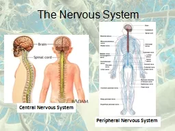
2. IntroductionBrain is that portion of the central nervous system which is contained with the cranium. One of the largest organs of the body, about 100 billion neurons & 10-50 trillion neuroglia make up the brain, which has a mass of about 1300 gm. or 1.3-1.4 kg. in adults.Because of the difference in average body weight of males & females, males have 10% larger brain than those of females (on average), however it is fact that there is no correlation between the brain size and intelligence. The brain and spinal cord develop from ectoderm arranged in a tubular structure called the neural tube. The anterior part of the neural tube expands and constricts into three regions called primary brain vesicles: prosencephalon (forebrain), mesencephalon (mid brain) & rhombencephalon (hindbrain).
3.
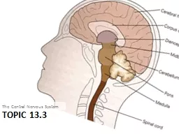
4. Parts of BrainBrain divided into following three major parts; Fore brain, Mid brain & Hind BrainForebrain divides into cerebrum & diencephalon.Midbrain divides into tectum & tegmentum.Hindbrain divides into pons, cerebellum & medulla oblangata.Note: Midbrain, Pons & Medulla oblangata are collectively termed as the brain stem.Brain stem is continuous with the spinal cord and consists Medulla oblangata, Pons & midbrain.Cerebellum is posterior to the brain stem.Diencephalon is superior to the brain stem and consists Thalamus, Hypothalamus & Epithalamus.Cerebrum is the largest part of brain, consist cerebral hemispheres which include cerebral cortex, white matter & basal nuclei.
5.
6. Coverings of the brainThe brain is protected by the cranial bones & the cranial meninges. The meninges are three layered tissues that completely surround the brain and spinal cord.The meninges that protect the brain are termed as cranial meninges which is present between the skull and brain.The meninges that protect spinal cord known as spinal meninges, present between the vertebral foramina & spinal cord.The cranial & spinal meninges have the same basic structure and same names which are; Dura mater, Pia mater & Arachnoid mater.The dura & arachnoid maters are separated by the subdural space, whereas the arachnoid and pia maters are separated by the arachnoid space that contains the cerebrospinal fluid.
7.
8. Coverings of the brainDura MaterOutermost covering of brain & spinal cord.In brain known as cerebral dura mater & in spinal cord known as spinal dura mater.The various parts of the brain are separated by three extensions of cerebral dura mater;Falx cerebri-separates two hemispheres of cerebrumFalx cerebelli- separates the two hemispheres of cerebellumTentorium cerebelli- separates the cerebrum from the cerebellumSpinal dura mater is continuous with cerebral dura mater and it forms a loose sheath around the spinal cord.There are some Dural folds in the spinal dura mater, which stabilize and support the spinal cord.
9. Coverings of the brainArachnoid materLayer of fibrous tissue that lies between the dura mater & pia mater.Separated from the dura mater by the subdural space and from the pia mater by the subarachnoid space containing cerebrospinal fluid.Pia materDelicate layer of connective tissue containing many minute blood vessels that adheres to the completely cover brain including dipping into each fissure.Thicker than arachnoid mater, specially over the spinal cord.
10. Blood brain barrierThe Blood-brain-barrier (BBB) protects brain cells from harmful substances and pathogens by preventing passage of many substances from blood into brain tissue.The BBB is established by;presence of tight junctions between the endothelial cells in the capillary walls, (b) presence of a thick basement membrane around the capillaries (decrease permeability) and (c) presence of the processes from astrocytes (neuroglial cells)The barrier exists in all parts of the brain except in some areas of the hypothalamus.The substance that can cross the barrier and enter the brain tissues are oxygen, carbon dioxide, water, glucose, amino acids, electrolytes and certain drugs that include sulfonamides, tetracyclines and many lipid soluble drugs.
11. Significance of BBBPrevents the entry of injurious substances & protein bounded substances in the brain.Maintains constant neuronal environment within the CNS by preventing the escape of neurotransmitters into the blood.Some metabolic enzymes are also present on BBB that inactivate some toxic metabolites and prevents any damage to the brain.Prevents adverse CNS effects of aqueous soluble drugs (e.g. Penicillin, Streptomycin & Thiopentone).Entry of hormones is also restricted, thereby preventing the disturbances of normal rhythm of the body.
12. Ventricles of BrainThe ventricles are the cavities within the brain filled with CSF which act as shock absorber for the CNS and circulates nutrients. Brain contains four irregular shaped cavities known as right lateral ventricle, left lateral ventricle, third ventricle & fourth ventricle.Lateral ventriclesThese cavities lie within the cerebral hemispheres, one on each side of median plane just below the corpus callosum. The right and left ventricles are separated from each other by a thin membrane, the septum pellucidum.Each lateral ventricle connects with the third ventricle by a narrow opening called intraventricular foramen.
13. Ventricles of BrainThird ventricleSituated below the lateral ventricles between the right & left halves of the thalamus. It connects with the fourth ventricle by a canal, known as the cerebral aqueduct.Fourth ventricleDiamond shaped cavity present below and behind the third ventricle, between the cerebellum and pons.Roof of Fourth ventricles contains three openings connects with the subarachnoid space of the brain & spinal meninges which allow the passage of CSF from the spinal cord, brain & other ventricles of brain.
14. Cerebrospinal FluidClear, colorless liquid that protect the brain & spinal cord against injuries by acting as a shock absorber.It carries oxygen, glucose and other required nutrients from the blood to neurons and neuroglia.This fluid continuously circulates through the subarachnoid space around the brain and spinal cord as well as their cavities.Site of CSF production are the choroid plexuses, which are the network of capillaries surrounded by ependymal cells (type of glial cells) in the linings of the ventricle walls.The CSF formed in the choroid plexuses of each lateral ventricle flows into the third ventricle through intraventricular foramen where more CSF is added by the choroid plexus present in the roof of the third ventricle. The fluid flows into the fourth ventricle through cerebral aqueduct where some more fluid is added by choroid plexus of the fourth ventricle.
15. Cerebrospinal FluidFrom the roof of fourth ventricle, CSF flows through foramina (openings) into the subarachnoid space and completely surrounds the brain and spinal cord.The movement of CSF occurs due to pulsation of blood vessels, respiration.CSF continuously secreted at a rate of about 0.5 ml/min. (720 ml/day) but the volume remains fairly constant at about 120 ml, which means that CSF reabsorbed as rapidly as it is formed by the choroid plexus.Functions of CSF1. Supports and protects the brain and spinal cord.2. Acts as a fluid buffer and protects the brain from shock.3. Medium for the interchange of substances such as nutrients and waste products between CSF and nerve cells.
16. Functions of Brain- Cerebruma. Cerebral hemispheres- A deep furrow divided the cerebrum into two halves i.e. Right hemisphere works creativity & Left hemispheres works for logic abilities.Corpus callosum (Bundles of axon) connects these two hemispheres.Each hemisphere of the cerebrum responsible for following functions;i. It is responsible for controlling all the voluntary activities.ii. Receive & process information pertaining to sensory stimuli.iii. Associated with functions like memory, intelligence, reasoning & learning.
17.
18. Functions of Brain- Cerebrumb. Cerebral Cortex- Three different type of activities related to cerebral cortex are divided into 4 lobes- Frontal, Parietal, Temporal & Occipital lobes;i. Mental activities- Related to memory, intelligence, thinking, reasoning, sense of responsibility, moral sense & learningii. Sensory perception- Related to perception of sensory stimuli including pain, temperature, touch, sight, hearing, taste and smell.iii. Voluntary control- Initiation & control over contraction of voluntary muscles.
19. Brain wavesAt any instant, brain neurons are generating millions of nerve impulses (action potentials). Taken together, these electrical signals are called brain waves. Brain waves generated by neurons close to the brain surface, mainly neurons in the cerebral cortex, can be detected by sensors called electrodes placed on the forehead and scalp. A record of such waves is called an electroencephalogram (electro- electricity; -gram recording) or EEG. Electroencephalograms are useful both in studying normal brain functions, such as changes that occur during sleep, and in diagnosing a variety of brain disorders, such as epilepsy, tumors, trauma, hematomas, metabolic abnormalities, sites of trauma, and degenerative diseases. The EEG is also utilized to determine if “life” is present, that is, to establish or confirm that brain death has occurred.
20. Brain waves1. Alpha waves: These rhythmic waves occur at a frequency of about 8–13 cycles per second. (The unit commonly used to express frequency is the hertz [Hz]. One hertz is one cycle per second.) Alpha waves are present in the EEGs of nearly all normal individuals when they are awake and resting with their eyes closed. These waves disappear entirely during sleep. 2. Beta waves: The frequency of these waves is between 14 and 30 Hz. Beta waves generally appear when the nervous system is active—that is, during periods of sensory input and mental activity. 3. Theta waves: These waves have frequencies of 4–7 Hz. Theta waves normally occur in children and adults experiencing emotional stress. They also occur in many disorders of the brain. 4. Delta waves: The frequency of these waves is 1–5 Hz. Delta waves occur during deep sleep in adults, but they are normal in awake infants. When produced by an awake adult, they indicate brain damage.