
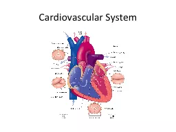
Introduction Cardiovascular system delivers oxygen and nutrients to cells of body tissue Heart muscular pump Blood vessels fuel line and transportation network Blood Vessels and the Circulation of Blood ID: 913546
Download Presentation The PPT/PDF document "Cardiovascular System Cardiovascular Sy..." is the property of its rightful owner. Permission is granted to download and print the materials on this web site for personal, non-commercial use only, and to display it on your personal computer provided you do not modify the materials and that you retain all copyright notices contained in the materials. By downloading content from our website, you accept the terms of this agreement.


Slide1
Cardiovascular System
Slide2Cardiovascular System:
Introduction
Cardiovascular system:
delivers oxygen and nutrients to cells of body tissue
Heart
(muscular pump)
Blood vessels
(fuel line and transportation network)
Slide3Blood Vessels and the
Circulation of Blood
Arteries
are the vessels that
lead away
from the heart.
Veins
have thinner walls than arteries and move
deoxygenated blood toward the heart
from the tissues.
Capillaries
are the smallest vessels. They form the point of
exchange for oxygen and nutrients into body cells
and waste products coming from body cells.
Slide4Blood Vessels
Slide5Blood Circulation/Systemic Circulation
Slide6Anatomy of the
Heart
Tricuspid valve
(cusps are flaps of the valves): between the
right atrium
and
right ventricle
Pulmonary valve:
between the
right ventricle
and
pulmonary artery
Mitral valve:
between the
left atrium
and
left ventricle
Aortic valve:
between the
left atrium
and
aorta
Slide7Pathway of Blood
through the Heart
Slide8Heartbeat and Heart Sounds
Two phases of the heartbeat:
D
iastole:
relaxation
S
ystole:
contraction
The diastole-systole cardiac cycle occurs between 70 to 80 times per minute (100,000 times per day).
The heart pumps 3 ounces of blood with each contraction. This means that about 5 quarts are pumped per minute (75 gallons an hour and about 2000 gallons a day).
Slide9Heart Sounds
Closure of valves associated with sounds
“
lubb-dubb
,
lubb-dubb
”
lubb
:
closure of the tricuspid and mitral valves
at the beginning of systole
dubb
:
closure of the aortic and pulmonary valves
at the end of systole
murmur
: an
abnormal heart sound caused by improper valve closure
Slide10Conduction System of the Heart
Sinoatrial
node (SA node)
: the
pacemaker
of the heart
Pacemaker
: origin of electrical impulse causing walls of the
atria to contract and force blood into the ventricles (ending diastole)
Slide11Conduction System of the
Heart
Atrioventricular
node (AV node): This sends the excitation wave to a bundle of specialized fibers called the
atrioventricular
bundle or Bundle of His.
Bundle of His (pronounced
“
hiss
”
):
Helps form conduction
myofibers
that extend to ventricle walls and stimulate them to contract, beginning systole. A short rest period follows.
The pacemaker begins wave of excitation again.
ECG or EKG
(electrocardiogram):
The record used
to detect electrical changes in heart muscle as the heart beats
.
Slide12Blood Pressure
Blood pressure:
The force that blood exerts on arterial walls
Expressed
as a fraction:
systolic pressure/
diastolic pressure
Example: 120/80 mm Hg
Hypertension
(high
blood pressure)
:
when BP > 140/90 mm Hg
Slide13Pathology:
the Heart and Blood Vessels
Heart
Arrhythmias (without normal heart rhythm)
Heart block (
atrioventricular
block)
Flutter
*
Fibrillation
AF: most common type of cardiac arrhythmia,
Electrical impulses move randomly throughout the atria, causing atria quiver instead of contracting a coordinated rhythm
.
VF: Electrical impulses move randomly throughout the
ventricles
. This life threatening situation may result in sudden
cardiac arrest
or death.
Slide14Pathology:
the Heart and Blood
Vessels
Heart
*Congenital heart disease (CHF)
:
The heart is
unable to pump the required amount of blood
.
In the U.S., primarily the result of
high blood pressure
and
coronary artery disease
(see next slide)
Results in
pulmonary
edema (fluid build up in lungs)
Fatal if untreated
Slide15Pathology:
the Heart and Blood
Vessels
Coronary artery disease
(CAD)
*
Atherosclerosis
- Deposition of fatty compounds of inner lining of coronary arteries.
*Thrombotic occlusion:
blockage of coronary artery by clot.
*Ischemia-
Blood flow is decreased or stopped completely, leads to necrosis.
*Necrosis:
D
eath of part of the myocardium.
*Infarction:
heart attack, and area of necrosis is known as infarct.
Slide16Pathology:
the Heart and Blood
Vessels
Coronary artery disease (CAD)
Surgical therapies for CAD
Coronary artery bypass grafting (CABG)
Percutaneous coronary intervention (PCI)
Slide17Pathology:
the Heart and Blood
Vessels
*
Endocarditis
- inflammation of inner lining of heart from bacteria.
*
Murmur-
extra heart sound, heard between normal beats.
*
Pericarditis:
inflammation of membrane (pericardium) surrounding the heart. Usually results from disease elsewhere in the body.
*
Aneurysm
- widening (dilation) of an arterial wall. Danger is an aneurysm can rupture and hemorrhage.
Slide18Pathology: Heart and Blood Vessels
*
Raynaud disease-
recurrent episodes of pallor and cyanosis primarily in fingers & toes. Intense constriction and vasospasms of arterioles often by young/healthy women. Idiopathic but triggered by; cold temps, stress, or smoking.
*
Varicose veins-
swollen/twisted veins caused by damaged valves that fail to prevent the backflow of blood. Blood collects in veins which makes them much larger in size. Thrombosis can occur. Hemorrhoids are varicose veins near anus.
Slide19Clinical Procedures: Treatment
Cardioversion
(defibrillation)
*
Endartectomy
: surgical removal of plaque from inner layer of an artery. Fatty deposits and
thromboses
are removed to open clogged arteries.
http://www.youtube.com/watch?v=5lZItV39v_Q
circulation
Heart transplantation
Thrombolytic therapy
Transcatheter
aortic valve replacement (TAVR)
Slide20Clinical Procedures: Treatment
*Coronary
artery bypass graft (CABG) surgery
A,
A section of a vein is removed from the leg and
anastomosed
to a coronary artery to bypass an area of arteriosclerotic blockage.
B,
An internal
mammary artery is grafted to a coronary artery
to bypass blockage.
://www.youtube.com/watch?v=3Nf6Q2skGOM
Slide21Treatment Procedures
*Percutaneous
coronary intervention (PCI)
Includes:
angioplasty
(PTCA), stent placement
, laser angioplasty, and
atherectomy
http://
www.youtube.com
/
watch?v
=N7nghr9TpSU