
98 Abstract Cold agglutinin diseaseCAgD is a type of autoimmune hemolytic anemia which generally occurs in adults and is characterized by the presence of IgM antibodies directed against polysaccha ID: 940803
Download Pdf The PPT/PDF document "Cold agglutinin disease in sepsis A" is the property of its rightful owner. Permission is granted to download and print the materials on this web site for personal, non-commercial use only, and to display it on your personal computer provided you do not modify the materials and that you retain all copyright notices contained in the materials. By downloading content from our website, you accept the terms of this agreement.


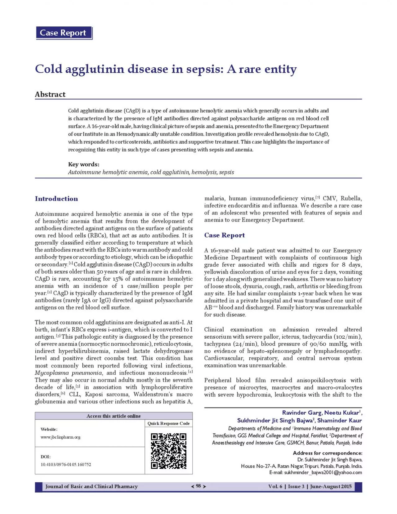
98 Cold agglutinin disease in sepsis: A Abstract Cold agglutinin disease(CAgD) is a type of autoimmune hemolytic anemia which generally occurs in adults and is characterized by the presence of IgM antibodies directed against polysaccharide antigens on red blood cell surface. A16-year-old male, having clinical picture of sepsis and anemia, presented to the Emergency Department of our )nstitut• in an (•modynamically unstabl• condition. )nv•stigation profil• r•v•al•d h•molysis du• to CAgD, which responded to corticosteroids, antibiotics and supportive treatment. This case highlights the importance of recognizing this entity in such type of cases presenting with sepsis and anemia. Key words: Autoimmune hemolytic anemia, cold agglutinin, hemolysis, sepsis Ravinder Garg, Neetu Kukar 1 , Sukhminder Jit Singh Bajwa 2 , Shaminder Kaur Departments of Medicine and 1 Immuno Haematology and Blood Transfusion, GGS Medical College and Hospital, Faridkot, 2 Department of Anaesthesiology and Intensive Care, GSMCH, Banur, Patiala, Punjab, India Address for correspondence: Dr. A, Ratan Nagar, Tripuri, Patiala, Punjab, India. sukhminder_bajwa2001@yahoo.com Introduction Autoimmune acquired hemolytic anemia is one of the type of hemolytic anemia that results from the development of antibodies directed against antigens on the surface of patients (RBCs), that act as auto antibodies. It is generally classified either according to temperature at which the antibodies react with the RBCs into warm antibody and cold antibody types or according to etiology, which can be idiopathic or secondary. [1] (CAgD) occurs in adults years of age and is rare in children. CAgD is rare, accounting for 15% of autoimmune hemolytic case/million people per year. [2] CAgD is typically characterized by the presence of IgM antigens on the red blood cell surface. The most common cold agglutinins are designated as anti-I. At birth, infant’s RBCs express i-antigen, which is converted to I antigen. [3] This pathologic entity is diagnosed by the presence of severe anemia(normocytic normochromic), reticulocytosis, indirect hyperbilirubinemia, raised lactate dehydrogenase level and positive direct coombs test. This condition has most commonly been reported following viral infections, Mycoplasma pneumonia , and infectious mononucleosis. [4] They may also occur in normal adults mostly in the seventh decade of life, [5] in association with lymphoproliferative disorders, [6] CLL, Kaposi sarcoma, Waldenstrom’s macro globunemia and various other infections such as hepatitis A, malaria, human immunodeficiency virus, [7] infective endocarditis and influenza. We describe a rare case of an adolescent who presented with features of sepsis and anemia to our Emergency Department. Case Report A 16-year-old male patient was admitted to our Emergency Medicine Department with complaints of continuous high grade fever associated with chills and rigors for 8 yellowish discoloration of urine and eyes for 2 day along with generalized weakness. There was no history of loose stools, dysuria, cough, rash, arthritis or bleeding from any site. He had similar complaints 1-year back when he was admitted in a private hospital and was transfused one unit of AB +ve blood and discharged. Family history was unremarkable for such disease. Clinical examination on admission revealed altered sensorium with severe pallor, icterus, tachycardia (24/min), blood pressure of 90/60 no evidence of hepato-spl
enomegaly or lymphadenopathy. Cardiovascular, respiratory, and central nervous system examination was unremarkable. Peripheral blood film revealed anisopoikilocytosis with presence of microcytes, macrocytes and macro-ovalocytes with severe hypochromia, leukocytosis with the shift to the Access this article online Website: www.jbclinpharm.org Quick Response Code DOI: 10.4103/0976-0105.160752 Case Report Journal of Basic and Clinical PharmacyVol. 6 | Issue 3 | June-August 2015 Vol. 6 | Issue 3 | June-August 2015Journal of Basic and Clinical Pharmacy 99 left, nucleated RBC’s 31/100 white blood cells while platelet count was normal. Coomb’s test showed positive DCT and weakly positive ICT [Tables 1 and 2]. Blood grouping was performed, but due to the presence of auto-agglutination in the sample at room temperature(~7–8°C), it was difficult to perform cell and serum grouping. The fresh blood sample in citrate vial was taken and incubated at 37°C for 30 Then, cell washing was done 8–10 saline at 37°C. 5% cell suspension of washed sample was prepared. Cross-match was done with AB +ve packed red blood (PRBCs) unit with tube method, and gel method and it was found to be compatible with patient’s serum. Then, the PRBCs unit was washed three times with warm normal saline and subsequently issued to the patient. The PRBCs unit was transfused when the temperature of the unit reached to about 37°C, that is, body temperature, so as to avoid any hemolytic reaction. Overall, three units of washed PRBCs of AB +ve blood group were transfused: 1 unit/day on alternate days. The chest X-ray and abdominal ultrasonography were unremarkable. In addition to blood component transfusions, he was administered corticosteroids, antibiotics and other supportive treatment and was discharged in a satisfactory condition. Discussion In the present case, anemia was most probably due to CAgD secondary to some infectious cause; however blood culture was negative. Serological test for detection of M. was not done. Cold agglutinins were confirmed by auto control positive test at 4°C and direct Coombs test Blood was transfused after 2days because of the difficulty in cross matching. Patient responded favorably to intravenous methylprednisolone and antibiotics. Corticosteroids and intravenous immunoglobulins are the mainstay of treatment though they are less effective in cold antibody mediated hemolysis, when the disease course is usually prolonged. Anti CD 20 molecule rituximab has been used in cold and warm agglutinin disease, the main disadvantage with their use being the possibility of flare-up of any underlying infection. [8] In 2005, a case of autoimmune hemolytic anemia was reported in a child having sickle cell disease who developed severe anemia induced by cold agglutinin hemolysis after Mycoplasma infection. Complete blood count falsely decreased RBC count and hematocrit and falsely RBC clumping at room temperature; this disappeared after warming at 37°C. Anti C3b-C3d was present on red cells, and indirect antiglobulin test revealed a circulating cold agglutinin. Furthermore, anti-Mycoplasma pneumoniae IgM antibody was detected in serum. Careful evaluation of CBCs and peripheral blood smears is required in cases of worsening anemia among sickle cell patients, and consideration should be given to cold hemagglutinin disease as an etiology. [9] In 2013, another case of cold agglutinin positive autoimmune hemolytic anemia was reported, which was d
iagnosed to be due to Klebsiella infection after ruling-out other causes that have been reported earlier. The patient continued to have hemolysis even after treatment of the underlying infection, intravenous methylprednisolone pulse and intravenous immunoglobulin. He responded to plasmapheresis with resolution of hemolysis. [10] The present case got admitted in an hemodynamicaly unstable condition with sepsis and although, unlike previous studies, no specific organism could be isolated but still the whole process of agglutination appears to be attributable to sepsis. An interesting feature of our case was that as the sepsis improved, Table Investigations Result Haemoglobin 2.0 g/dl TLC 39.73/cumm Differential leukocyte count 7, metamyelocytes 12, polymorphs eosinophils PLT count 13×10 3 /uL Reticulocyte count % 2.1 Total serum bilirubin 5.6 mg/dl Indirect bilirubin 4.7 mg/dl LDH 278.7 IU/L Malaria card test Negative Urine examination (routine Normal PTI % 87.5 INR 1.1 HBsAganti Nonreactive LDH: Lactate dehydrogenase, PTI: Prothrombin time index, INR: surface antigen, HCV: Anti hepatitis C virus, HIV: Anti human immunodeficiency virus, TLC: Total leukocyte count, PLT: Platelet Table 1 Investigation 29.12.2013 06.01.2014 Hb 2.0 g/dl 10.5 g/dl TLC 39.73×10 3 13.79×10 3 PLT 13×10 3 /uL 70×10 3 /uL MCV 155 fL 102 fL RBS 99 mg/dl Blood urea 37 mg/dl 19 mg/dl Serum creatinine 0.9 mg/dl 0.4 mg/dl Serum uric acid 11.2 mg/dl 5.8 mg/dl Total bilirubin 5.6 mg/dl 1.6 mg/dl Conjugated bilirubin 0.9 mg/dl 0.3 mg/dl Unconjugated bilirubin 4.7 mg/dl 1.3 mg/dl SGOT 44 IU/L 48 IU/L SGPT 18 IU/L 42 IU/L ALP 159 247 LDH 278.7 IU/L RBS: Random blood sugar, LDH: Lactate dehydrogenase, SGOT: Serum glutamic oxaloacetic transaminase, SGPT: Serum glutamic transaminase, ALP: Alkaline phosphatase, TLC:leukocyte count, PLT: Platelet, MCV: Mean corpuscular volume Garg, et .: Cold agglu�nin in sepsis 100 the agglutination process declined and only then it was possible to specify the blood group and give a blood transfusion. Conclusion The present case highlights the importance of recognizing CAgD in patients presenting in a hemodynamically unstable condition with sepsis and severe anemia. References F, ChestermanB. de Gruchy’s Clinical Haematology in Medical Practice. 6 th ed. New Delhi: Wiley; 2013. p. E, Langholm W, et Primary chronic cold agglutinin disease: A population based clinical study of 86 DM, Rodberg Blood Banking and Transfusion Practices. 6 th ed. New Delhi: Jaypee; 2013. p. MA. Cold agglutinin disease and cryoglobulinemia. Clin Lymphoma 2005;5:290 C. Agewise distribution of idiopathic cold agglutinin disease. Z S, WanJY, HanrahanLR. Future development of lymphoproliferative disorders in patients with autoimmune hemolytic anemia. Clin Cancer Res 2001;7:791 MW. HIVassociated autoimmune hemolytic anemia: an update. AIDS Patient Care STDS 2001;15:217 P, BrethonP, LandmanF, A. Treatment of childhood autoimmune haemolytic anaemia with rituximab. Lancet 2001;358:1511 TL, LasaterOE, WangWC. Acase of hemoglobin SC disease with cold agglutinininduced hemolysis. Am J Hematol 2005;78:37 10. E, YadavTP. Klebsiella infectionassociated autoimmune Indian Acad Clin Med 2013;14:190 How to cite this article: Garg R, Kukar N, Bajwa SJ, Kaur S. Cold agglutinin disease in sepsis: A rare entity. J Basic Clin Pharma 2015;6:98-100. Source of Support: Garg, et .: Cold agglu�nin in sepsis Journal of Basic and Clinical PharmacyVol. 6 | Issue 3 | June-August 201