
Smith R Muehlenbachs A Schaenmann J Baxi S Koo S Blau D et al Three Cases of Neurologic Syndrome Caused by DonorDerived Microsporidiosis Emerg Infect Dis 2017233387395 httpsdoiorg103201eid2303161580 ID: 1009558
Download Presentation The PPT/PDF document "Figure 2 Figure 2. Transmission electron..." is the property of its rightful owner. Permission is granted to download and print the materials on this web site for personal, non-commercial use only, and to display it on your personal computer provided you do not modify the materials and that you retain all copyright notices contained in the materials. By downloading content from our website, you accept the terms of this agreement.

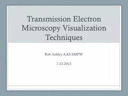


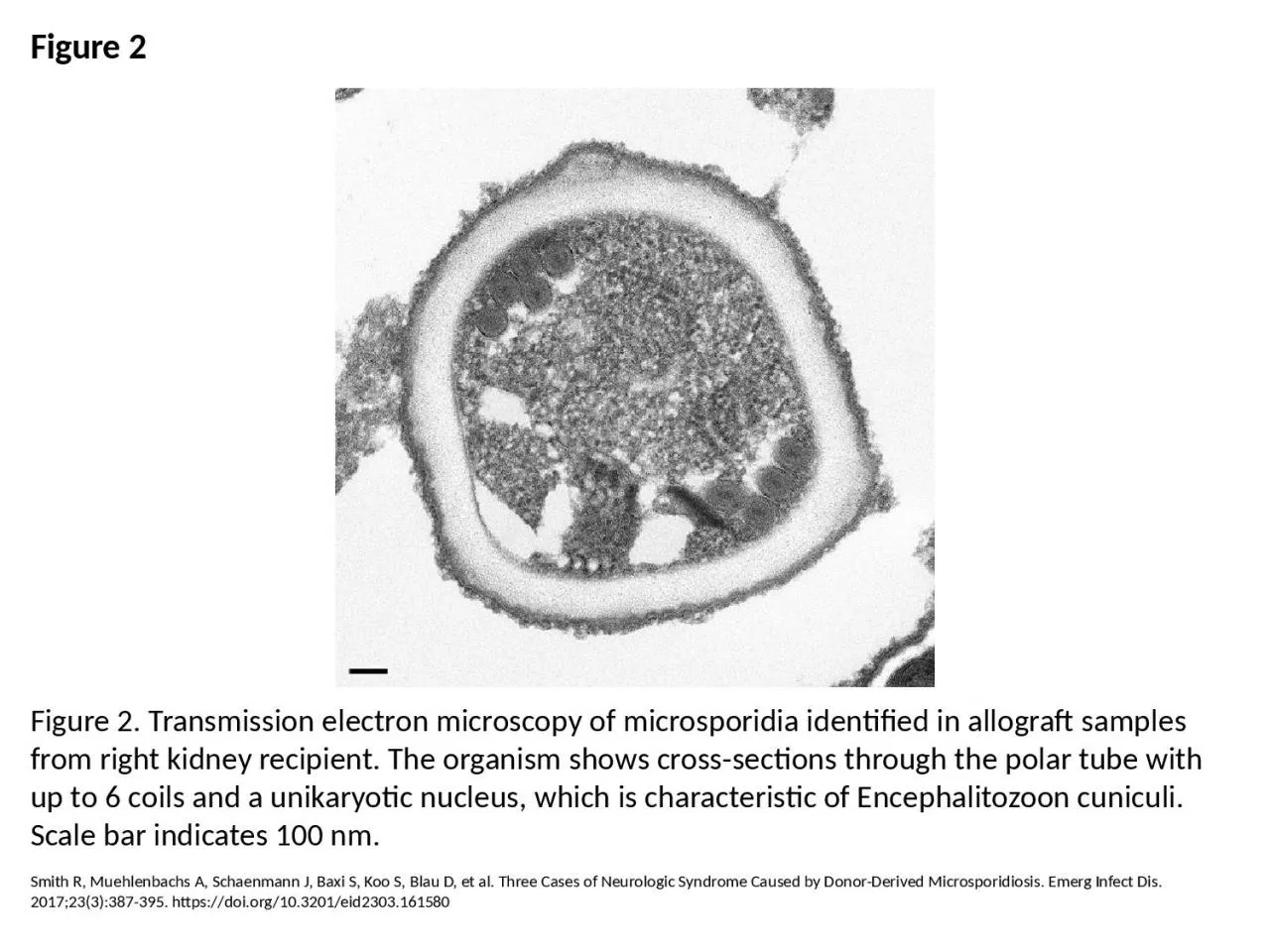
1. Figure 2Figure 2. Transmission electron microscopy of microsporidia identified in allograft samples from right kidney recipient. The organism shows cross-sections through the polar tube with up to 6 coils and a unikaryotic nucleus, which is characteristic of Encephalitozoon cuniculi. Scale bar indicates 100 nm.Smith R, Muehlenbachs A, Schaenmann J, Baxi S, Koo S, Blau D, et al. Three Cases of Neurologic Syndrome Caused by Donor-Derived Microsporidiosis. Emerg Infect Dis. 2017;23(3):387-395. https://doi.org/10.3201/eid2303.161580