
Basavaraju SV Kuehnert MJ Zaki S Sejvar J Encephalitis Caused by Pathogens Transmitted through Organ Transplants United States 20022013 Emerg Infect Dis 201420914431451 httpsdoiorg103201eid2009131332 ID: 1039104
Download Presentation The PPT/PDF document "Figure 4 Figure 4. Brain images showing ..." is the property of its rightful owner. Permission is granted to download and print the materials on this web site for personal, non-commercial use only, and to display it on your personal computer provided you do not modify the materials and that you retain all copyright notices contained in the materials. By downloading content from our website, you accept the terms of this agreement.


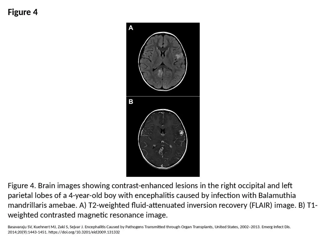
1. Figure 4Figure 4. Brain images showing contrast-enhanced lesions in the right occipital and left parietal lobes of a 4-year-old boy with encephalitis caused by infection with Balamuthia mandrillaris amebae. A) T2-weighted fluid-attenuated inversion recovery (FLAIR) image. B) T1-weighted contrasted magnetic resonance image.Basavaraju SV, Kuehnert MJ, Zaki S, Sejvar J. Encephalitis Caused by Pathogens Transmitted through Organ Transplants, United States, 2002–2013. Emerg Infect Dis. 2014;20(9):1443-1451. https://doi.org/10.3201/eid2009.131332