
A Neurosurgical perspective Dominic Thompson Department of Paediatric Neurosurgery Great Ormond Street London Flemish 1515 Anon Am J Med Genetics Cervical spinal pathology in Downs syndrome ID: 911505
Download Presentation The PPT/PDF document "Management of Cervical Spine Disease in ..." is the property of its rightful owner. Permission is granted to download and print the materials on this web site for personal, non-commercial use only, and to display it on your personal computer provided you do not modify the materials and that you retain all copyright notices contained in the materials. By downloading content from our website, you accept the terms of this agreement.


Slide1
Management of Cervical Spine Disease in Down's Syndrome: A Neurosurgical perspective
Dominic ThompsonDepartment of Paediatric NeurosurgeryGreat Ormond StreetLondon
Slide2Flemish (1515)
Anon
Am J Med Genetics
Slide3Cervical spinal pathology in Downs syndrome-The controversies
AetiologyDiagnosisScreeningTreatment
Slide4Aetiology
Slide5AetiologyPrimary features
Ligamentous laxityHypotoniaHypermobility
Slide6AetiologyPrimary features
Ligamentous laxityHypotoniaHypermobilitySkeletal consequencesCraniovertebral instability
Scoliosis
Hip instability
Patellar instability
Slide7Cervical spinal pathology in Downs syndrome
Occipito-atlantal subluxationAtlantoaxial subluxationOs OdontoideumIncomplete C1 ringCervical stenosisSegmentation anomaly
Premature cervical spondylarthrotic myelopathy
Slide8Cervical spinal pathology in Downs syndrome
Occipito-atlantal subluxationAtlantoaxial subluxationOs OdontoideumIncomplete C1 ringCervical stenosis
Segmentation anomaly
Premature cervical spondylarthrotic myelopathy
Slide9Os Odontoideum – congenital or acquired?
C1
OS
Slide10Os odontoideum – developmental anatomy
Sclerotomes cranial and caudal moieties – scleromites Cranial – hypocentrum - disc Caudal – centrum - body
Slide11Os odontoideum – developmental anatomy
Sclerotomes cranial and caudal moieties – scleromites
Cranial – hypocentrum - disc
Caudal – centrum - body
Slide12Os odontoideum – developmental anatomy
Sclerotomes cranial and caudal moieties – scleromites
Cranial – hypocentrum - disc
Caudal – centrum - body
C1 hypocentrum
C1 centrum
Slide13Os odontoideum – developmental anatomy
Sclerotomes cranial and caudal moieties – scleromites
Cranial – hypocentrum - disc
Caudal – centrum - body
C2 hypocentrum -regresses
C2 centrum
Slide14Os odontoideum – developmental anatomy
Congenital theory of the Os.
= Failure of incorporation of the C1 centrum
Implications
Large os (entire C1 centrum)
Base below joint
Slide15Os odontoideum – developmental anatomy
Congenital theory of the Os.
= Failure of incorporation of the C1 centrum
Absence of C1 centrum rare
Slide16Os odontoideum – developmental anatomy
Acquired theory
Trauma to
odontoid
due to instability
Implications
Os relatively small
Base at or above joint line
Slide17Os odontoideum – developmental anatomy
Slide18Radiological findings in 12 symptomatic cases (Nader-Sepathi et al 2005
)Atlanto-dental interval 5-13mm (av.7mm)Neural canal width 6-11mm (av.8mm)Atlanto-occipital instability 2 casesOs odontoideum 1
0
cases
Posterior spina bifida 3 cases
Anterior spina bifida 2 casesPars defect C4 1 case
Slide19Radiological findings in 12 symptomatic cases (Nader-Sepathi et al 2005
)Atlanto-dental interval 5-13mm (av.7mm)Neural canal width 6-11mm (av.8mm)Atlanto-occipital instability 2 casesOs odontoideum 1
0
cases
Posterior spina bifida 3 cases
Anterior spina bifida 2 casesPars defect C4 1 case
Slide20Atlanto-dental interval 5-13mm (av.7mm)Neural canal width 6-11mm (av.8mm)
Atlanto-occipital instability 2 casesOs odontoideum 10 casesPosterior spina bifida 3 casesAnterior spina bifida 2 casesPars defect C4 1 case
Radiological findings in 12 symptomatic cases (
Nader-Sepathi et al 2005
)
Slide21Craniovertebral instability
Os OdontoideumDelayed Atlantal ossification
Slide22Diagnosis
Slide23Diagnosis of craniovertebral instabilityin Downs Syndrome
Aims To identify children with or at risk of spinal cord damage due to craniovertebral instabilityTo instigate effective treatment
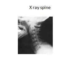
Slide24What to measure?
Slide25Defining atlantoaxial subluxation
Atlanto-dental interval (ADI) NR <5mmNeural canal width (NCW) NR >14mm
ADI
Slide26Defining atlantoaxial subluxation
Atlanto-dental interval (ADI) NR <5mmNeural canal width (NCW) NR >14mm
NCW
Slide27Defining atlantoaxial subluxation in Downs syndrome
Author
N
ADI
Pueschel 1986
404
4.5mm
Morton 1995
90
4mm
Semine
85
4.5mm
Elliot 1988
67
4mm
Van Dyke 1988
34
5mm
Slide28Defining atlantoaxial subluxation in Downs syndrome
Author
N
ADI
%
Pueschel 1986
404
4.5mm
14.6
Morton 1995
90
4mm
7.8
Semine
85
4.5mm
12
Elliot 1988
67
4mm
10
Van Dyke 1988
34
5mm
9
Slide29Defining atlantoaxial subluxation in Downs syndrome
Author
N
ADI
%
Symptoms
Pueschel 1986
404
4.5mm
14.6
1.5
Morton 1995
90
4mm
7.8
0
Semine
85
4.5mm
12
1.2
Elliot 1988
67
4mm
10
0
Van Dyke 1988
34
5mm
9
0
Slide30Acute vs chronic deteriorationNader-Sepathi et al 20055/12 acute deterioration
Preceeding symptoms in 4Davidson 198831 Downs with neurological symptoms/signs29 symptoms for >1 mnth
Slide3112 years old
Downs syndromeGait deteriorationClinical signs of myelopathy
Slide32Extension
Flexion
Slide33ADI Changes over time
Morton et al 1995
N=67
Measurements 5 years apart
Slide34Poor clinical and radiological correlation
Ferguson 1987
Slide35Poor clinical and radiological correlationGait abnormality as a measure of AAS
Specificity 81%Sensitivity 50%
Gait
Radiographs
Normal Abnormal
Total
Normal
Abnormal
92 (91) 9 (9)
21 (70) 9 (30)
101
30
Selby 1991
Slide36Poor clinical and radiological correlationGait abnormality as a measure of AAS
Specificity 81%Sensitivity 50%Poor reproducibilty in repeat films
Gait
Radiographs
Normal Abnormal
Total
Normal
Abnormal
92 (91) 9 (9)
21 (70) 9 (30)
101
30
Selby 1991
Slide37Defining Occipito-atlantal subluxation
Coexisting occipito- atlantal instability is common 40-50%Treadwell 1990
Taggart 2000
Difficult to diagnose
Slide38Defining Occipito-atlantal subluxation
Powers ratio
Slide39Defining Occipito-atlantal subluxation
Basion-Axial IntervalHarris et al
Slide40Defining Occipito-atlantal subluxation
Wiesel et al
Slide41Defining Occipito-atlantal subluxation“Measurement of atlanto-occipital translation by any of these methods is not reproducible”
Lori et al 1996
Slide42Limitations of Radiographic Measurements in Downs Syndrome
ADI No consensus on measurementPoor reproducibilityPoor correlation with symptomsO-C instability, present but difficult to measure
Slide43Screening
Slide44Should Children With Down Syndrome be Screened for Atlantoaxial Instability?
Slide45Should Children With Down Syndrome be Screened for Atlantoaxial Instability?Special Olympics 1983
All patients with Downs syndrome should have lateral cervical spine X rays ADI>4.5 mm mandates exclusion from high risk sporting activitiesAmerican Academy of Pediatrics 1984Department of Health Standing Medical Advisory Committee 1986
Slide46Should Children With Down Syndrome be Screened for Atlantoaxial Instability?Special Olympics 1983
All patients with Downs syndrome should have lateral cervical spine X rays ADI>4.5 mm mandates exclusion from high risk sporting activitiesAmerican Academy of Pediatrics 1984Department of Health Standing Medical Advisory Committee 1986
Slide47Screening Incidence must be common
Screening test should be sensitive and specificAsymptomatic AAS must be a risk factor for symptomatic AASA safe and effective intervention should be available
Slide48British Guidelines 2009Downs Medical Interest Group of UK
,
Cervical spine X rays are not recommended for the asymptomatic child with Downs syndrome
Medical personnel and carers need to be aware of the potential for neurological symptoms to develop
Slide49British Guidelines 2009Downs Medical Interest Group of UK
Warning Signs
Neck pain,
Abnormal head posture,
Torticollis, (Wry neck)
Reduced neck movements,
Deterioration of gait and/or frequent falls
Increasing fatigability on walking,
Deterioration of manipulative skills,
Slide50British Guidelines 2009Downs Medical Interest Group of UK
Action
Full physical and neurological examination
Specialist Referral
Slide51British Gymnastics AssociationGuidelines for the participation of individuals with Downs syndrome in gymnastics and trampolining
For all applicants to: Registered clubsCompetitive eventsScreening by medical practitionerApproved byBritish Gymnastics Medical Commission
Gymnastics and movement for people with disabilities Technical Committee
Slide52Clinical screening criteria
A. No signs of myelopathyIncreasing muscle weakness, alteration in muscle toneLoss of sensationDecreasing co-ordinationDiminishing kinaesthetic awarenessChange in walking pattern
New incontinence
B. Neck flexion
Flex to chin on chest without difficulty
C. Neck muscle control
Pulled to sitting position maintaining head neck alignment
Slide53Results1995-2009
399 applications from Individuals with Down syndrome
6 positive responses
4 Poor head neck muscle control
2 Flex to chin on chest without difficulty
No further follow up
Slide54Results393 granted membership
No injuries referable to the cervical spine reported during the time period3 in National Squad
Slide55Treatment
Slide56Treatment“A safe an effective intervention should be available”
Slide57Surgery for AAS in Down Syndrome
Author
Number
Re-op (%)
Neurol. Deterioration
Osseous fusion
Doyle
15
6 (40)
4 (27)
12 (80)
Thompson
15
6 (40)
1 (8)
15 (100)
Segal
10
3 (30)
2 (20)
(both died)
4 (40)
Taggard
25
0
0
23 (96)
Slide58Reasons for failureN=15, 6 failures
Inadequate immobilisationInadequate reduction Inadequate fixationInadequate age
Slide59Inadequate immobilisation
Reduce and immobilise prior to surgery Maintain immobility post operatively in non instrumented fixation
Slide60Inadequate fixation
Non union associated
Resorption of rib graft
Limited C1-C2
Nader-Sepathi et al 2005
Doyle et al 1996
Slide61Inadequate reduction5 yearsAttempted posterior fixation
Inadequate reduction Construct failure
Slide62Stage I
MUA, halo body jacketStage II
Transoral odontoidectomy
Stage II
Posterior decompression and revision of instrumentation
Slide63Case 1: Downs syndrome
Stage IMUA, halo body jacketStage II
Transoral odontoidectomy
Stage II
Posterior decompression and revision of instrumentation
Slide64Case 1: Downs syndrome
Stage IMUA, halo body jacketStage II
Transoral odontoidectomy
Stage II
Posterior decompression and revision of instrumentation
Slide65Inadequate reduction
Transoral decompression6 out of 15 casesAll had os odontoideum
Slide66Slide67Slide68ConclusionsOsseous anomalies at the CCJ may be consequence rather than cause of instability
Morphometric measures of AAI and OCI lack sufficient sensitivity and specificity to justify screeningEducation rather than screening
Slide69ConclusionsEarly investigation and treatment of symptomatic patients
Beware the child with Downs!EnsureEffective immobilisationAdequate reductionRigid fixation
Slide70