
Cardiovascular toxicities include arrhythmias accelerated atherosclerosis myocardial infarction and heart failure 1 Steroid use has been previously associated with nonischemic cardiomyopathy a potentially lifethreatening cardiotoxicity as an adverse effect of using exogenous glucocorticoids ID: 1033330
Download Presentation The PPT/PDF document "Self-administered supraphysiologic doses..." is the property of its rightful owner. Permission is granted to download and print the materials on this web site for personal, non-commercial use only, and to display it on your personal computer provided you do not modify the materials and that you retain all copyright notices contained in the materials. By downloading content from our website, you accept the terms of this agreement.


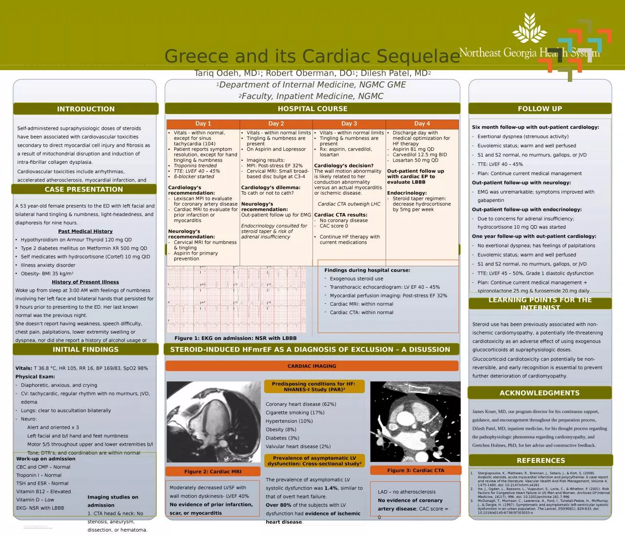
1. Self-administered supraphysiologic doses of steroids have been associated with cardiovascular toxicities secondary to direct myocardial cell injury and fibrosis as a result of mitochondrial disruption and induction of intra-fibrillar collagen dysplasia.Cardiovascular toxicities include arrhythmias, accelerated atherosclerosis, myocardial infarction, and heart failure.1Steroid use has been previously associated with non-ischemic cardiomyopathy, a potentially life-threatening cardiotoxicity as an adverse effect of using exogenous glucocorticoids at supraphysiologic doses. Glucocorticoid cardiotoxicity can potentially be non-reversible, and early recognition is essential to prevent further deterioration of cardiomyopathy. James Kruer, MD, our program director for his continuous support, guidance, and encouragement throughout the preparation process, Dilesh Patel, MD, inpatient medicine, for his thought process regarding the pathophysiologic phenomena regarding cardiomyopathy, and Gretchen Holmes, PhD, for her advise and constructive feedback.Greece and its Cardiac SequelaeTariq Odeh, MD1; Robert Oberman, DO1; Dilesh Patel, MD21Department of Internal Medicine, NGMC GME 2Faculty, Inpatient Medicine, NGMC Six month follow-up with out-patient cardiology:Exertional dyspnea (strenuous activity)Euvolemic status; warm and well perfused S1 and S2 normal, no murmurs, gallops, or JVDTTE: LVEF 40 – 45% Plan: Continue current medical management Out-patient follow-up with neurology:EMG was unremarkable; symptoms improved with gabapentinOut-patient follow-up with endocrinology:Due to concerns for adrenal insufficiency; hydrocortisone 10 mg QD was started One year follow-up with out-patient cardiology:No exertional dyspnea; has feelings of palpitations Euvolemic status; warm and well perfused S1 and S2 normal, no murmurs, gallops, or JVDTTE: LVEF 45 – 50%, Grade 1 diastolic dysfunction Plan: Continue current medical management + spironolactone 25 mg & furosemide 20 mg daily CASE PRESENTATIONA 53 year-old female presents to the ED with left facial and bilateral hand tingling & numbness, light-headedness, and diaphoresis for nine hours.Past Medical HistoryHypothyroidism on Armour Thyroid 120 mg QDType 2 diabetes mellitus on Metformin XR 500 mg QDSelf medicates with hydrocortisone (Cortef) 10 mg QIDIllness anxiety disorderObesity- BMI 35 kg/m2History of Present IllnessWoke up from sleep at 3:00 AM with feelings of numbness involving her left face and bilateral hands that persisted for 9 hours prior to presenting to the ED. Her last known normal was the previous night.She doesn’t report having weakness, speech difficulty, chest pain, palpitations, lower extremity swelling or dyspnea, nor did she report a history of alcohol usage or other recreational drug usage. INTRODUCTIONHOSPITAL COURSEEVIDENCE OF ASYMPTOMATIC LV DYSFUNCTION VS HEART FAILUREFOLLOW UPLEARNING POINTS FOR THE INTERNISTACKNOWLEDGMENTSREFERENCESINITIAL FINDINGSVitals: T 36.8 °C, HR 105, RR 16, BP 169/83, SpO2 98% Physical Exam: Diaphoretic, anxious, and crying CV: tachycardic, regular rhythm with no murmurs, JVD, edemaLungs: clear to auscultation bilaterallyNeuro: Alert and oriented x 3 Left facial and b/l hand and feet numbness Motor 5/5 throughout upper and lower extremities b/l Tone, DTR’s, and coordination are within normal Figure 1: EKG on admission: NSR with LBBB Work-up on admissionCBC and CMP – NormalTroponin I – Normal TSH and ESR - Normal Vitamin B12 – Elevated Vitamin D – Low EKG- NSR with LBBB Imaging studies on admission1. CTA head & neck: No stenosis, aneurysm, dissection, or hematoma.2. MRI brain with contrast: No evidence of recent infarction or acute intracranial process.STEROID-INDUCED HFmrEF AS A DIAGNOSIS OF EXCLUSION – A DISUSSION Coronary heart disease (62%)Cigarette smoking (17%) Hypertension (10%) Obesity (8%) Diabetes (3%) Valvular heart disease (2%)CARDIAC IMAGINGPredisposing conditions for HF: NHANES-I Study (PAR)2Moderately decreased LVSF with wall motion dyskinesis- LVEF 40%No evidence of prior infarction, scar, or myocarditisLAD – no atherosclerosisNo evidence of coronary artery disease; CAC score = 0 Day 1Day 2Day 3Day 4Vitals - within normal, except for sinus tachycardia (104)Patient reports symptom resolution, except for hand tingling & numbnessTroponins trendedTTE: LVEF 40 – 45%ß-blocker startedCardiology’s recommendation:Lexiscan MPI to evaluate for coronary artery diseaseCardiac MRI to evaluate for prior infarction or myocarditis Neurology’s recommendation:Cervical MRI for numbness & tingling Aspirin for primary preventionVitals - within normal limitsTingling & numbness are presentOn Aspirin and Lopressor Imaging results:MPI: Post-stress EF 32% Cervical MRI: Small broad-based disc bulge at C3-4Cardiology’s dilemma:To cath or not to cath? Neurology’s recommendation:Out-patient follow up for EMGEndocrinology consulted for steroid taper & risk of adrenal insufficiencyVitals - within normal limitsTingling & numbness are presentRx: aspirin, carvedilol, losartanCardiology’s decision?The wall motion abnormality is likely related to her conduction abnormality versus an actual myocarditis or ischemic disease. Cardiac CTA outweigh LHCCardiac CTA results:No coronary diseaseCAC score 0Continue HF therapy with current medications Discharge day with medical optimization for HF therapyAspirin 81 mg QDCarvedilol 12.5 mg BIDLosartan 50 mg QDOut-patient follow up with cardiac EP to evaluate LBBBEndocrinology: Steroid taper regimen: decrease hydrocortisone by 5mg per weekStergiopoulos, K., Mathews, R., Brennan, J., Setaro, J., & Kort, S. (2008). Anabolic steroids, acute myocardial infarction and polycythemia: A case report and review of the literature. Vascular Health And Risk Management, Volume 4, 1475-1480. doi: 10.2147/vhrm.s4261He, J., Ogden, L., Bazzano, L., Vupputuri, S., Loria, C., & Whelton, P. (2001). Risk Factors for Congestive Heart Failure in US Men and Women. Archives Of Internal Medicine, 161(7), 996. doi: 10.1001/archinte.161.7.996McDonagh, T., Morrison, C., Lawrence, A., Ford, I., Tunstall-Pedoe, H., McMurray, J., & Dargie, H. (1997). Symptomatic and asymptomatic left-ventricular systolic dysfunction in an urban population. The Lancet, 350(9081), 829-833. doi: 10.1016/s0140-6736(97)03033-xFigure 2: Cardiac MRIFigure 3: Cardiac CTAPrevalence of asymptomatic LV dysfunction: Cross-sectional study3The prevalence of asymptomatic LV systolic dysfunction was 1.4%, similar to that of overt heart failure.Over 80% of the subjects with LV dysfunction had evidence of ischemic heart disease.Findings during hospital course:Exogenous steroid useTransthoracic echocardiogram: LV EF 40 – 45% Myocardial perfusion imaging: Post-stress EF 32% Cardiac MRI: within normalCardiac CTA: within normal