
zebrafish retina Kartik Sreepada 1 Xiangping Chu 1 Salvatore L Stella Jr 2 1 Department of Basic Medical Sciences UMKC School of Medicine Kansas City MO United States 2 Department of Neural and Behavioral Sciences Penn State College of Medicine Hershey PA United States ID: 1014581
Download Presentation The PPT/PDF document "The effects of illumination of VNUT expr..." is the property of its rightful owner. Permission is granted to download and print the materials on this web site for personal, non-commercial use only, and to display it on your personal computer provided you do not modify the materials and that you retain all copyright notices contained in the materials. By downloading content from our website, you accept the terms of this agreement.


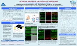
1. The effects of illumination of VNUT expression in zebrafish retinaKartik Sreepada1, Xiangping Chu1, Salvatore L Stella Jr.21Department of Basic Medical Sciences, UMKC School of Medicine, Kansas City, MO, United States2Department of Neural and Behavioral Sciences, Penn State College of Medicine, Hershey, PA, United StatesINTRODUCTIONAdenosine, a breakdown product of ATP, suppresses photoreceptor transmitter release in the outer retina by modulating input from photoreceptors onto second-order neurons.1–3 The discovery of the vesicular nucleotide transporter (VNUT) for ATP supports a vesicular release of ATP.4,5VNUT is expressed both in inner and outer zebrafish retina.We hypothesized that VNUT expression in the outer zebrafish retina is regulated by illumination.METHODSFollowing IACUC approval, zebrafish 6 months or older were euthanized with tricaine 1X at either 7 am (night) or 7 pm (day). See Figure 2. Vertical retinal sections were prepared by immersing zebrafish eyes in 4% PFA for 15 minutes followed by overnight cryoprotection in 30% sucrose. The eyecups were embedded in optimal cutting temperature (OCT) compound and cut on a cryostat (12 microns)Indirect immunofluorescence using antibodies for zebrafish VNUT was usedAll images were acquired sequentially to avoid spectral bleed-through on an upright fixed stage confocal microscope.RESULTSCONCLUSIONVNUT is expressed in the outer retina within the OPL.VNUT is not expressed at terminals of photoreceptors but localized to postsynaptic (i.e. second-order) neurons (e.g. horizontal cells).VNUT is also present in the IPL, however we have not characterized all cell types expressing VNUT in the inner retinaInterestingly, VNUT is highly expressed at the end-feet of Müller cells, suggesting that ATP is released by the glial cells in the inner retina through a vesicular mechanism.These findings are compelling and need to be explored further in order to fully elucidate the impact purines have on neurotransmission in vertebrate retina.Future studies: compare night and daytime conditions on VNUT expression, use western blotting to assess VNUT protein levels in the retinas of day-primed and night-primed zebrafish, and map the expression of VNUT in outer and inner retinal neurons during these conditions.CREDITS/DISCLOSURE/REFERENCESStella et al (2003). Endogenous adenosine reduces glutamatergic output from rods through activation of A2-like adenosine receptors. J. Neurophysiol. 90(1):165-74.Stella et al (2007). Adenosine inhibits voltage-dependent Ca2+ influx in cone photoreceptor terminals of the tiger salamander. J. Neurophysiol. 85(1):1126-37.Stella et al. (2009). Adenosine suppresses exocytosis from cone terminals of the salamander retina. Neuroreport. 20(1):923-29.Sawada et al. (2008). Identification of a vesicular nucleotide transporter. Proc Natl Acad Sci. 105:5683-6.Wall & Dale. (2008). Activity-dependent release of adenosine: a critical re-evaluation of mechanism. Curr Neuropharmacol. 6: 329-37.I would like to thank the UMKC Sarah Morrison Fellowship for providing a grant to support my research efforts on this project Figure 2. Experimental strategy. This diagram shows a 24 hour clock. The zebrafish are on a 12 hr light/dark cycle with the day beginning at 8 a.m. and ending at 8 p.m., animals were collected at 7 a.m. (yellow) near the completion of the dark cycle, and at 7 p.m. (yellow), near the completion of the light cycle. Tissues were collected for immunocytochemistry (VNUT localization).Figure 3. Zebrafish retina immunostained with anti-VNUT antibody (green) upper panel, and VNUT preadsoprtion (lower panels). Scale bars are 10 microns.Antibodies (host)SourceDilutionSlc17a9 (VNUT) T-12 (rabbit)Santa Cruz Biotech1:1000ZPR-1 (mouse)ZIRC (Univ. of Oregon)1:2000GFAP (mouse)UC Davis NeuroMab1:500 to 1:1000Figure 1. Conversion of ATP into adenosine. Ecto-NTPDases convert ATP to ADP and AMP, and the 5’-ectonucleotidase converts AMP to adenosine.Figure 4. Zebrafish retina immunostained with anti-VNUT antibody (green) and anti-ZPR antibody (red). Inset illustrates a zoom of the OPL as shown in the box under the MERGE panel. Arrows indicate terminals of zebrafish photoreceptors. Scale bars are 10 micronsFigure 5. Zebrafish retina immunostained with anti-VNUT antibody (green) and anti-GFAP antibody (red). Arrows indicate Muller cell endfeet. Scale bars are 10 microns.2. Daytime expression: ZPR-1 does not co-localize with VNUT in photoreceptors.3. Daytime expression: GFAP positive Müller cell endfeet co-localize with VNUT immunoreactivity in the inner1. VNUT expression in the zebrafish retinaA. SLC17A9/VNUT is a vesicular transporter that concentrates ATP into synaptic vesicles. B. Preadsorption of VNUT immunoreactivity with VNUT peptide blocks specific labeling in zebrafish retina proteinVNUTVNUT + DICVNUT + Block PepBlock + DICOPLINLIPLGCLPLOPLINLIPLGCLPLVNUTZPR-1MERGEOPLINLIPLGCLPLOPLVNUTGFAPMERGEOPLINLIPLGCLPLFigure Key: PL=Photorceptor Layer, OPL= Outer Plexiform Layer, INL= Inner Nuclear Layer, IPL= Inner Plexiform Layer, GCL= Ganglion Cell Layer