
Limb Mgr Veronika Mrkvicová physiotherapist Examination Methods in Rehabilitation 26102020 Nerves of the Upper Limb Axillary nerve Musculocutaneuous nerve ID: 913040
Download Presentation The PPT/PDF document "Nerves of the Upper" is the property of its rightful owner. Permission is granted to download and print the materials on this web site for personal, non-commercial use only, and to display it on your personal computer provided you do not modify the materials and that you retain all copyright notices contained in the materials. By downloading content from our website, you accept the terms of this agreement.


Slide1
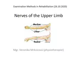
Nerves of the Upper Limb
Mgr. Veronika Mrkvicová (physiotherapist)
Examination Methods in Rehabilitation (26.10.2020)
Slide2Nerves of the
Upper LimbAxillary nerve
Musculocutaneuous nerveRadial nerveMedian nerve
Ulnar
nerve
Slide3Brachial plexusNetworking of spinal
nerves, formed by ventral (anterior rami) of
cervical spinal nerves C5-C8 and thoracic spinal nerves T1It
is
responsible
for
cutaneous
(
sensory
)
and
muscular
(motor)
innervation
of
the
entire
upper
limb
Slide4Brachial plexus5 main
nerves arise from brachial plexus:
Axillary nerveMusculocutaneuous nerveRadial nerveMedian
nerve
Ulnar
nerve
Slide5Nerves of the Upper Limb - sensitivity
Slide6Slide7The cutaneous inervation of the right hand
Slide8Axillary nerveFrom root C5-C6
Arise from posterior cord of brachial plexus
at the level of axilla
Slide9Slide10Innervations of the Axillary nerve
Muscular innervations:Anterior branch –
anterior and lateral fiber of deltoid muscles
Posterior
branch
–
teres
minor
and
posterior
fiber
of
deltoid
Cutaneuos
innervation
:
Superior
lateral
brachial
cutaneous
nerve
Carry
information
from
the
shoulder
joint
Skin
covering
inferior
region
of
deltoid
muscles
Slide11Axillary nerve paralysis
Frequently injured due to shoulder dislocation because
of the proximity of this joint
Paralysis
of
the
deltoid
and
teres
minor
–
it
results
in
weakness
of
the
arm abduction
Slide12Musculocutaneous nerve
Slide13Innervation of the Musculocutaneous
nerve
Slide14Slide15Median nerve
The median nerve is one of the 5 main nerves originating from the brachial plexusIt originates from the lateral and medial cords of the brachial plexus, and has contributions from ventral roots of
C5 and C6 (lateral cord) and C8 and
T
h
1
(medial cord)
The median nerve is the only nerve that passes through the
carpal tunnel
Slide16Median nerveorigin:lateral root - lateral cord of the brachial plexus
medial root - medial cord of the brachial cordcourse: laterally to the axillary artery, descends in the arm between biceps brachii and triceps brachii muscles, courses through the forearm with the ulna nerve and vessels before entering the carpal tunnel to the hand
major branches: anterior interosseous nerve, palmar cutaneous branch, motor branch in the handmotor supply: flexor compartment of the fore
arm
,
thenar
and intrinsic hand muscles
sensory supply:
palmar
aspect of the thumb, index, middle and radial half of the ring fingers
Slide17Median nerve – branches:
anterior interosseous nerve supplies all the flexor muscles of the forearm
apart from flexor carpi ulnaris and the ulnar half of flexor
digitorum
profundus
motor branch in the hand
- supplies
thenar
muscles
and the radial two
lumbricals
palmar
cutaneous
branch
-
cutaneous
innervation
to the
palmar
aspect of the thumb, index and middle fingers and the radial half of the ring finger
articular branches to the elbow, wrist, carpal and phalangeal joints
Slide18Slide19Median nerve palsy
ape-hand deformity
Slide20Median nerve palsy – signs and
symptoms:Lack of ability to abduct and oppose the thumb due to paralysis of the thenar muscles
. This is called "ape-hand deformity„Sensory loss in the thumb, index finger, long finger, and the radial aspect of the ring fingerWeakness in forearm
pronation
and wrist and finger flexion
Difficulties
in
Activities of daily living
(ADL)
such as brushing teeth, tying shoes, making phone calls, turning door knobs and writing
Slide21Median nerve palsy – causes:
deep, penetrating injuries to the arm, forearm, or wristor blunt force trauma or neuropathyCan be separated into 2 subsections
- high and low MNP:High MNP involves lesions at the elbow and forearm areasLow MNP results from lesions at the wristc
ompression
at the different levels of the
median nerve
produce variable symptoms and/or syndromes
, t
he areas are:
Underneath
Struthers' ligament
Passing by the
bicipital
aponeurosis
(also known as
lacertus
fibrosus
)
Between the two heads of the
pronator
teres
Compression in the carpal tunnel causes
carpal tunnel syndrome
Slide22Tests of median nerve function
Thumb „circles“
Thumb
opposition
Thumb
flexion
Fingers
flexion
Slide23Slide24Carpal tunnel syndrome
Slide25Ulnar nerve
It originates from the C8-T1 nerve roots (and occasionally carries C7
fibres) which form part of the medial cord of the brachial plexus, and descends on the posteromedial aspect of the humerus
Slide26Ulnar nerve – motor inervation
In the forearm, via the muscular branches
of ulnar nerve: Flexor carpi
ulnaris
Flexor
digitorum
profundus
(
medial
half)
In
the
hand
, via
the
deep
branch
of
ulnar
nerve:
hypothenar muscles Opponens
digiti
minimi
Abductor
digiti
minimi
Flexor
digiti
minimi
brevis
The
third
and
fourth
lumbrical
muscles
Dorsal
interossei
Palmar
interossei
Adductor
Pollicis
Flexor
pollicis
brevis
(
deep
head
)
In
the
hand
, via
the
superficial
branch
of
ulnar
nerve
:
Palmaris
brevis
Slide27Ulnar nerve – sensory inervation
Sensory inervation to the V.digit and the medial half of the IV.digit, and the corresponding part of the palm:
Palmar
branch of
ulnar
nerve
:
cutaneous
innervation
to the anterior skin and nails
Dorsal
cutaneous
branch of
ulnar
nerve
:
cutaneous
innervation
to the dorsal medial hand and the dorsum of the medial 1.5 fingers
Slide28Slide29Ulnar nerve palsyThe
ulnar nerve can suffer injury anywhere between its proximal origin of the brachial plexus all the way to its distal branches in the handIt is the most commonly injured nerve around the elbowAlthough it can be damaged under various circumstances, it is commonly injured by local trauma or physical
impigement ("pinched nerve")Injury of the ulnar nerve at different levels causes specific motor and sensory deficits
Slide30Ulnar nerve palsy – position of
the hand
An ulnar claw (or
claw hand
, or
´
Spinster's Claw
´)
The
metacarpophalangeal
joints
of the 4th and 5th fingers are extended
and the
Interphalangeal
joints are flexed
,
thumb
IP
flexion
Slide31Ulnar nerve palsyThe hand will show hyper-extension of the MCP and flexion of the distal and proximal IP joints of the 4th and 5th digits
The clawing will become most obvious when the person is asked to flex the digits from an extended position as the 4th and 5th digits can not flex 1st, 2nd and 3rd digits will partially flex giving them a "claw-like" appearance, this happens because the Thenar muscles (Abductor
pollicis brevis, Flexor Pollicis brevis and Opponens pollicis) are innervated by the median nerve as the first two
lumbricals
of digit 2 and 3 are
Slide32Froments´ test (Froments´ sign)
Tests for the action of adductor pollicis A patient is asked to hold a flat object
(a piece of paper), between their thumb and index finger (pinch grip)The examiner then attempts to pull the object out of the subject's handsA normal individual will be able to maintain a hold on the object without difficultyWith
ulnar
nerve palsy, the patient will experience difficulty maintaining a hold and will compensate by flexing the FPL (flexor
pollicis
longus
) of the thumb to maintain grip pressure causing a pinching effect
Slide33Ulnar nerve palsy – fingers abduction
Unability to spread (abduct) or pull together (adduct) the fingers against resistance (because the ulnar
nerve innervates the palmar and dorsal interossei of the hand)
Slide34Ulnar nerve palsy – muscles atrophy
Patients with this deficit will become increasingly easy to identify over time as the paralyzed first dorsal interosseous muscle atrophies, leaving a prominent hollowing between the thumb and forefinger
Slide35Ulnar entrapmentIt
is a condition where the ulnar nerve becomes trapped or pinched due to some physiological abnormalitiesIt is classified by location
of entrapmentThe ulnar nerve passes through several small tunnels and outlets through the medial upper extremity, and at these points the nerve is vulnerable to compression or entrapment - a so-called "pinched nerve„
The nerve is particularly vulnerable to injury when there has been
a disruption in the normal anatomy
Slide36Ulnar entrapment
It can be classified by specific local
causes, including:Problems originating at the
neck
:
thoracic
outlet
sy
,
cervical
spine
pathology
,
tight
anterior
scalene
muscles
Problems
originating in the
chest: tight pectoralis minor muscles
Brachial
plexus
abnormalities
Elbow
pathology
:
fractures
,
growth
plate
injuries
,
cubital
tunnel
sy
,
flexorpronator
aponeurosis
,
arcade
of
Struthers
Forearm
pathology
:
tight
flexor
carpi
ulnaris musclesWrist pathology: fractures, ulnar tunnel sy,
hypothenar
hammer
sy
Slide37Radial nerve
The radial nerve supplies the posterior portion of the upper limbIt innervates the medial and lateral heads of the triceps
brachii muscle of the arm, as well as all 12 muscles in the posterior osteofascial compartment of the forearm and the associated joints and overlying skin
It originates from the
brachial plexus
, carrying fibers from the ventral roots of spinal nerves C5, C6, C7, C8 & T
h
1
Slide38Radial nerveorigin: one of the two posterior cords of the
brachial plexuscourse: posteromedially with the axillary vessels, behind the humerus, then
anteriorly towards the elbow where it divides into superficial and deep branchesterminal branches: posterior interosseous (deep) and
superficial
radial
nerve
motor
:
wrist
and
finger
extension
sensory
: dorsal aspect of the thumb, index and middle fingers
Slide39Radial nerve – branches
muscular twigs in the arm – triceps brachii and anconeus muscles
superficial branch - supplies cutaneous sensation to the dorsal aspect of the hand and dorsal aspect of the first to third digits and the dorsal lateral aspect of the fourth fingerdeep branch - posterior
interosseous
nerve - extensor muscles in the forearm as well as
brachioradialis
articular
twigs
to the elbow and wrist joints
Slide40Slide41Slide42Radial nerve injury
The radial nerve is often injured in its
course close to the humerus, either from fracture or
pressure
from
direct
blow
to
the
humerus (
incorrect
use
of
a
crutch
)
Triceps
usually
escapes
because derivation of the
nerve giving off high in arm,
but
total
paralysis
of
the
extensor
of
the
wrist
and
digits
leads
to
the
dropped
wrist
deformities
Slide43Radial nerve palsy
Drop hand
Slide44Tests for extensors
Thumb extensors
Wrist
extensors
Slide45Brachial plexus injury
Slide46Erb´s Duchenne Palsy
Slide47Klumpke´s Palsy
Slide48Literature, e-sources
http://criticalcaremcqs.com/tag/aipgmee-mcqs/page/15/www.graysanatomyonline.com https://en.wikipedia.org/wiki/Median_nerve_palsy
http://www.slideshare.net/hermizan84/peripheral-nerves-of
-
upper
-limb?
related
=1
https://meded.ucsd.edu/clinicalmed/neuro2.htm
http://accessphysiotherapy.mhmedical.com/data/Multimedia/grandRounds/brachial/media/brachial_print.html
http://www.
slideshare.net
/hermizan84/
peripheral
-
nerves
-
of
-
upper
-limb?
related
=1
Slide49Thank you for your attention