
In 2D DIGE protein samples are labeled with size and chargematched CyDyefluorsThree fluorescently labeled protein samples can be combined and resolved using 2D gel electrophoresis isoelectric focus ID: 831513
Download Pdf The PPT/PDF document "Pooled internalStandard label with Cy2Pr..." is the property of its rightful owner. Permission is granted to download and print the materials on this web site for personal, non-commercial use only, and to display it on your personal computer provided you do not modify the materials and that you retain all copyright notices contained in the materials. By downloading content from our website, you accept the terms of this agreement.


Pooled internalStandard label with Cy2Pr
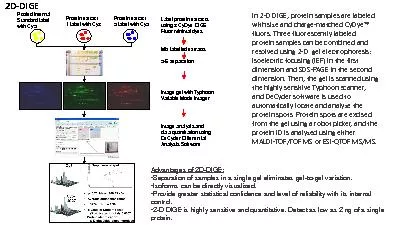
Pooled internalStandard label with Cy2Protein extract 1 label with Cy3Protein extract 2 label with Cy5Label protein extracts using 3 CyDyeDIGE Fluor minimal dyesMix labelled extractsE separationImage gel with Typhoon Variable Mode ImagerImage analysis and data quantitation using DeCyderDifferential Analysis SoftwareIn 2D DIGE, protein samples are labeled with size and chargematched CyDyefluorsThree fluorescently labeled protein samples can be combined and resolved using 2D gel electrophoresis: isoelectric focusing (IEF) in the first dimension and SDSPAGE in the second dimension.Then, the gel is scanned using the highly sensitive Typhoon scanner, and DeCydersoftware is used to automatically locate and analyze the protein spots. Protein spots are excised from the gel using a robot picker, and the protein ID is analyzed using either MALDITOF/TOF MS or ESIQTOF MS/MS.Advantages of 2DDIGE: Separation of samples in a single gel eliminates gelgel variation.Isoforms can be directly visualized.Provide greater statistical confidence and level of reliability with its internal control.D DIGE is highly sensitive and quantitative. Detect as low as 2 ng of a single protein. 2D-DIGE