
Bombay Hospital Journal Vol 51 No 1 2009127more common in females and is in all agegroup except childrenMortalityMorbidity Patients may beasymptomatic experience painful recurrenterosions de ID: 941425
Download Pdf The PPT/PDF document "126Bombay Hospital Journal Vol 51 No 1 2..." is the property of its rightful owner. Permission is granted to download and print the materials on this web site for personal, non-commercial use only, and to display it on your personal computer provided you do not modify the materials and that you retain all copyright notices contained in the materials. By downloading content from our website, you accept the terms of this agreement.


126Bombay Hospital Journal, Vol. 51, No. 1, 2009Corneal Epithelial Basement Membrane DystrophyDarshana B Rathod, Anjali D Nicholson, Bhavisha BidAbstractA 38 year old female presented with recurrent redness, pain, watering in both eyes since last 6-7 yrs having taken antibiotics, steroids, lubricating drops, anti-viral (acivir), hypersol, (ban-dage contact lenses) BCL for treatment with no positive family history. Her best correctedvisual acuity were 6/9 in both eyes for distance and N/6 for near with right eye showing lidoedema and conjunctival congestion. Corneal sensations and Schirmer’s test were normal.acuity were 6/9 in both eyes for distance and N/6 fornear with right eye showing lid oedema andconjunctival congestion. Corneal sensations andSchirmer’s test were normal.On examination, slit lamp findings suggested ofan oval corneal epithelial defect measuring 3 mm x 1mm, with positive fluoroscein staining and inferiorlimbal vascularisation in the right eye and left eyeshowing sub-epithelial map and dot pattern opacities.Patient was treated locally with mild steroid-fluromethanole and lubricating eye drops. SystemicDoxycycline 100 mg BD was prescribed for 6 weeks.PathophysiologyCorneal abnormalities associated withmap-dot-fingerprint dystrophy are the resultof a faulty basement membrane, which isthickened, multilaminar, and misdirected intothe epithelium. Maps histologically representareas of multilaminar basement membrane,which extend into the epithelium. Dots areintraepithelial microcysts that containnuclear, cytoplasmic, and lipid debris.Fingerprints are curvilinear clusters ofreduplicated and thickened basementmembrane and fibrillogranular material.Blebs, a less common manifestation arelocalized areas of fibrillogranular material orthickened basement membrane.3: In the US prevalence of map-dot-fingerprint dystrophy range from 2-43%of the general population and is found to beOphthalmology Department, B Y L Nair CharitableMunicipal Hospital, Mumbai 400 008.Introductionorneal map-dot-fingerprint dystrophy isby far the most common cornealdystrophy and is named from the appearanceof its characteristic slit lamp findings. Map-dot-fingerprint dystrophy also is known asepithelial basement membrane dystrophy,and Cogan microcystic epithelial dystrophy.It usually is classified as a dystrophy but fitsmore accurately into the cornealdegeneration category.Corneal dystrophies usually arehereditary, bilateral, progressive, and notassociated with systemic or local disease.1Map-
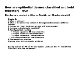
dot-fingerprint dystrophy has been foundin several families with a presumedautosomal dominant pattern, but in mostcases, it is not familial. It is not progressivebut rather variable and fluctuating in itscourse. Usually, it is bilateral but can beunilateral or very asymmetric inpresentation.Case ReportA 38 year old female presented with recurrentredness, pain, watering in both eyes since last 6-7 yrshaving taken antibiotics, steroids, lubricating drops,anti-viral (acivir), hypersol, BCL for treatment withno positive family history. Her best corrected visual Bombay Hospital Journal, Vol. 51, No. 1, 2009127more common in females and is in all agegroup except children.Mortality/Morbidity: Patients may beasymptomatic, experience painful recurrenterosions, decreased vision, or both.Clinical RelevanceRefraction is uncertain due to irregularastigmatism.Slit lamp findings includes the following:Corneal maps seen as irregular geographicshape, faint gray-white patches that maycontain clear oval areas. They vary greatlyin size (usually 1 mm to several mm) and areseen best with broad oblique illumination.Corneal dots seen as gray-white, puttylikeopacities, which can be round, comma-shaped,or irregular. They usually are 0.05-1.0 mmin size.Corneal fingerprints seen as clusters ofcontoured concentric lines 0.25-4.0 mm long.They are seen best with retroillumination.Corneal blebs are clear, round, bubble likedefects 0.05-0.2 mm in diameter. They areseen best with retroillumination.Keratometry shows irregular astigmatism.A placido disk or keratometer oftendemonstrates irregularity better thancomputed tomography.Conditions of elevated intraocular pressureand corneal decomposition which gives riseto corneal epithelial oedema mimickingcorneal pseudo fingerprints or shift lines cancause diagnostic dilemma.TreatmentMedical CareHypertonic drops or ointment help bothirregular astigmatism and recurrent cornealerosion problems. Sodium chloride (5%) dropfour times a day and ointment at bedtime is Fig. 1 : Oblique illumination showing corneal subepithelial map and dot dystrophy. Fig. 2 : Retro illumination view of basementmembrane dystrophy. 128Bombay Hospital Journal, Vol. 51, No. 1, 2009recommended. lubricating drops orointment is also preferred as it is found thatthere is no difference between hypertonic andnonhypertonic ointment.Capsule Doxycycline (100 mg) BD for sixweeks helps in the adhesion between cells ofepithelial basement membrane, mechanismof which is not known.Patching is done in case of acut
e episodesof corneal erosions.Bandage extended wear soft contact lensis useful but risk of infectious keratitis makesthis a secondary choice.Hard or gas-permeable contact lens is usedto improve vision by masking cornealirregular astigmatism but is poorly toleratedbecause of increased corneal fragility/erosionproblems.Surgical CareDebridement/superficial keratectomy isdone in case of significant visual loss fromassociated irregular astigmatism andrecurrent corneal erosions.Diamond burr superficial keratectomy:After epithelial debridement a diamond-dusted burr is used to polish the basementmembrane.Excimer laser phototherapeutickeratectomy is an excellent treatment forrecurrent corneal erosions associated withmap-dot-fingerprint dystrophy.4Corneal anterior stromal needle puncture:This procedure is not as successful forrecurrent erosions associated with map-dot-fingerprint dystrophy, which is usually morediffuse and often migratory.PreventionLubricating hypertonic saline or blandointment at bedtime is most of the timehelpful to prevent recurrent erosions.ComplicationsRecurrent erosions predispose the corneato infection.PrognosisMap-dot-fingerprint dystrophy findingsfluctuate but tend not to progress over time.Majority of patients are able to maintainsufficient vision and comfort for reading,driving, and other visual tasks, except duringepisodes of corneal erosions.Special ConcernsPatient with this dystrophy may bebothered by painful recurrent erosionepisodes and or decreased vision but are mostfrustrated by the unpredictability of thecondition.Map-dot -fingerprint dystrophy is a relativecontraindication for refractive procedures,such as LASIK or LASEK. Trauma from themicrokeratome sliding over the epithelialsurface or from flap manipulation is morelikely to occur in patients with map-dot-fingerprint dystrophy because of the poorlyadherent epithelium. Epithelial sloughing canlead to epithelial ingrowth and stromal melts.Surface ablation [PRK]) may be a betterrefractive procedure option for these patients.ReferencesDinh R, Rapuano CJ, Cohen EJ, et al. Recurrenceof corneal dystrophy after excimer laserphototherapeutic. Keratectomy. Ophthalmology1999; 106 : 1490-97.2.Bron AJ. Genetics of the corneal dystrophies:What have we learned in the past twenty-fiveyears. Cornea.2000; 19 : 699-711.3.Klintworth GK. Advances in the moleculargenetics of corneal dystrophies. Am JOphthalmology 1999; 128 : 747-54.4.External Disease and cornea-section 8.AmericanAcademy of Ophthalmology.2006-2007; 311-1