
Proteins Carbohydrates Nucleic acid Polysaccharides Peptides Amino acids Oligosaccharides Nucleosides Organic acids Small anions and cat ions of body fluids Principle of E lectrophoresis ID: 921304
Download Presentation The PPT/PDF document "Electrophoresis Electrophoresis is one o..." is the property of its rightful owner. Permission is granted to download and print the materials on this web site for personal, non-commercial use only, and to display it on your personal computer provided you do not modify the materials and that you retain all copyright notices contained in the materials. By downloading content from our website, you accept the terms of this agreement.


Slide1
Electrophoresis
Slide2Electrophoresis is one of the most important method for separating colloidal particles and biological molecules such as -
Proteins
Carbohydrates
Nucleic acid
Polysaccharides
Peptides
Amino acids
Oligosaccharides
Nucleosides
Organic acids
Small anions and cat ions of body fluids .
Slide3Principle of E
lectrophoresis
Electrophoresis is a separation technique.
Use to separate out ionizable substances in a sample.Based on the principle that – A charged particle in solution will migrate towards one of the electrodes when placed in an electrical field .
Slide4-
Cations
, positively charged particles will migrate to cathode.-Anions , negatively charged particles will migrate to anode.
Slide5The proteins found in plasma (TSP) all have amino acids as their subunits.
So ,each protein has its own specific isoelectric point.
Because of their different isoelectric points, each protein will move at a different rate when placed in an electrical field.Proteins with similar isoelectric points will migrate to a similar area in an electrical field
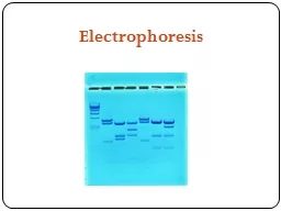
Slide6Serum protein electrophoresis
Hydragel – agarose gel
Serum proteins are separated into 6 groups:
Albuminα1 - globulinsα2 - globulins
β
1 - globulins
β
2 - globulins
γ
- globulins
Slide7Electrophoresis
Slide8Process of separation
When a voltage is applied across the electrodes, it generates a potential gradient.
This potential gradient apply a force on charge bearing molecules.
And drive a charged molecule towards an electrode.A frictional resistance oppose this movement of charged molecules .
Slide9which depends on size, shape of molecules, pore size of medium and viscosity of the buffer.
so, when a voltage is applied , molecules with different overall charges will begin to separate if they have different molecular sizes , shape and weight because experience different frictional forces.
Slide10Factors affect the rate of migration
The mobility or rate of migration of ions in electrophoresis depends upon-
Net charge of the moleculeSize and shape of the moleculeSupport medium propertiesStrength of the electrical field
Ionic strength of the bufferTemperature
Slide11Separation of sample
1. According to charge
2. According to size 3. According to shape
Slide12According to charge
1. When charged molecules are placed in an electric field, they migrate toward either the positive (anode) or negative (cathode) pole according to their charge.
Slide132 According to size
Smaller molecules run faster through the medium than larger ones.
3. According to shapeRound shape molecules have less frictional and electrostatic retardation than other shape molecules. so, moves fast in comparison to other shape molecules.
Slide14The Basic Components of electrophoresis
Electrophoresis chamber
-
is where it all process takes place. a. vertical b. horizontral 2. Electric power supply- provides the electricity that carries the molecules through the gel.Buffer solution - Buffer is chosen in that way that effective separation take place.Support medium – supporting medium is selected to ensure accurate and effective separation.
Slide15Electrophoresis unit
Slide16Electrophoresis unit,
available for running either –
Vertical
Slide17Horizontal gel electrophoresis unit
Slide18Type of electrophoresis
Vertical electrophoresis-
used for the seperation of proteins in polyacrylamide gelHorizontal electrophoresis – used for immunoelectrophoreis
, isolectric focusing, and for DNA , RNA in agarose gel.
Slide191. Power supply
The power supply is a source of constant voltage or current that provides energy to the electrodes.
This drives the movement of the ions in the medium and results in the movement and separation of the molecules or solutes in the specimen.
Slide202. Buffers
The two important purposes of the buffer are -
a. To create the pH
b. And to conduct the current.Main features of buffer are-Buffer maintain a constant state of ionization of the molecules being separated.The pH set by the buffer determines the net charge on the solutes.the resulting net charge determines which electrode the solutes migrate toward.The buffer ions carry the current during electrophoresis.
Slide21Besides setting the pH, the buffer also maintains the pH throughout the electrophoresis of the sample and control the conductivity of the gel.
In pH 8 - 9, proteins will take on a negative charge and migrate to the anode.
Most protein electrophoresis is performed at pH 8.6.
Slide22Tris
buffer (hydroxy methyl amino methane)- is a popular buffer for protein electrophoresis . It fix the pH at 8.6TBE buffer (Tris, Borate and EDTA) is often used for DNA fragments.
To prevent microbial growth and contamination, buffers must be refrigerated when not in use. Using a buffer at refrigerator temperature also improves band resolution and lessens evaporation during electrophoresis.
Slide233.
Support Medium-
For electrophoretic separation of solutes, the sample is placed on a support medium–It may be a- paper in paper electrophoresis and gel in gel electrophoresis
which is in contact with buffer for separation.
Slide24kinds of support medium commonly used are :
Starch
Agar/
agaroseCellulose acetatepolyacrylamide gel (PAGE) Paper (cellulose)
Slide25Properties of gel
-
The gel is a polymer whose composition and porosity is chosen based on the specific weight and composition of the sample to be analyzed.Agarose , and polyacrylamide gels are cross-linked, sponge like structure.
It is important that the support media is electrically neutral.
Slide26Cellulose acetate gel –
Cellulose is chemically reacted with acetic
anyhdride to form a cellulose acetate gel.Agarose gel is more often used than cellulose acetate gel for clinical electrophoresis.
The gel pores allow for separation of proteins based on their charge and mass.
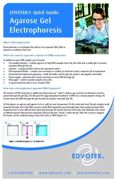
Slide27Agarose gel
A chain of sugar molecules
Extracted from seaweed
Slide28Agarose gel
For the separation of
(1) large protein or protein complex polynucleotide 50-30,000 base-pairs.The pore size is determined by adjusting the concentration of agarose in a gel (normally in the rank of 0.4-4%)
Common clinical uses of agarose gel electrophoresis (AGE) are separations of plasma proteins, hemoglobin variants, lipoproteins, and isoenzymes.
Slide29Agarose gel preparation
Slide30Gels solution are pored in the gel plate.
A comb with teeth is suspended in the gel until it cools.
When the gel has cooled and solidified, pulls the comb out of the gel.The teeth of the comb leave impressions in the gel called wells where the sample will be loaded.The gel plate is then placed in the electrophoresis chamber.Gel acts like a bridge through which the molecules must travel to get from one side of the chamber to the next.
Slide31Polyacrylamide Gels
Polyacrylamide electrophoresis (PAGE) is performed on a gel formed by polymerizing and cross-linking
acrylamides. These gels are stronger than agarose gels and also thermostable and transparent. In addition to separating fragments by charge and mass
, also separates solutes by molecular size.
Slide32Polyacrylamide gel
Slide33Types of electrophoresis
Lect-2
Slide341. Zone electrophoresis
Paper electrophoresis - - Protein is easily denatured due to high absorbance of filter paper. -Works for small peptides and amino acid separating.B
. Thin layer electrophoresis (TLE)C. Gel electrophoresis a. Starch gel electrophoresis b. Polyacrylamide gel electrophoresis (PAGE) c. Agarose gel electrophoresis
Slide35Slide36Separation of proteins by paper electrophoresis
Procedure -
In alkaline medium-Proteins are negatively charged.They will migrate towards anode in an electric field.Separation is based on net charge and difference in their molecular weight, their motilities are different.
Slide37Procedure
Immerse a cellulose paper strip into the electrophoresis buffer.
Take it out and evaporate water by drying this paper strip.Add 3-5 ul of sample on the strip 2 cm far from cathode side electrode.Put it on
electrophoretic device and connect it and electrode with the current device.
Slide38It is carried out at a constant voltage of 80V for 50-60 mints.
After
seperation process, take it out and immersed into fixer solution and than into a dye for 1 mints.Than put it into a destaining solution for rinsing.Observe the colour and width of the band on the strip to calculate the result.
Slide39Slide40Slide41Gel electrophoresis
Slide42Slide43Electroendosmosis
With a pH 8.0-9.0 used for protein electrophoresis, proteins take on a negative charge, that is a negative ion cloud forms.
As the negative ion cloud migrates to the anode, the proteins are pulled to the anode. Several gels used routinely for protein electrophoresis, attract positive ions from the buffer and form a positive ion cloud.
Slide44This ion cloud moves in the opposite direction to the cathode.
This phenomenon is called electro endosmosis or endosmosis.
The tension created by these oppositely moving ion clouds can affect the movement of sample macromolecules.
Slide45The migration of some proteins can be slowed, some proteins can become immobile, and other proteins are pushed toward the cathode.
The gamma globulin band in serum, urine, and other body fluids will separate more sharply by being pushed to the cathode.
Slide46Endosmosis
Slide47Other important types of electrophoresis
2.
Isoelectrric focusing3. Peptide mapping
4. Western blotting/immunostaining5. Immunoelectrophoresis6. Capillary electrophoresis7. Pulse field gel electrophoresis8. Isotachophoresis
Slide48Clinical applications of electrophoresis
Specific
protein
electrophoresis is used for quantitative analysis of serum proteins .Various classes of Hb can be separated by using this technique. It can be used in separation and quantization of major lipoproteins and determination of separate lipid profile .
Slide49It is being used for the separation of whole
chromosome
.Study of CSF protein can be done by electrophoresis.It can also be used for analysis of
Iso enzyme which are early diagnosis of various disorders of the body. Exampls are - Myocardial infarction, heart attack, liver disorders, muscular dystrophy and various type of cancer.
Slide50Creatine
kinase (CK), Lactate dehydrogenase (LDH) which is early signal of medical disaster can be estimated by electrophoresis. It can be used in diagnosis Multiple myeloma,
Liver cirrhosis, Immunodeficiency. In addition to that, we can do Western blotting for conformation of AIDS and Southern blotting for prenatal diagnosis of inborn errors, viral infection.