
It is one of the most serious emergency situation Early diagnosis and followed restoration of airflow is essential to prevent cardiac arrest or irreversible brain damage that occurs within minutes of complete airway obstruction ID: 908427
Download Presentation The PPT/PDF document "Upper airway obstruction" is the property of its rightful owner. Permission is granted to download and print the materials on this web site for personal, non-commercial use only, and to display it on your personal computer provided you do not modify the materials and that you retain all copyright notices contained in the materials. By downloading content from our website, you accept the terms of this agreement.


Slide1
Upper airway obstruction
Slide2It is one of the most serious emergency situation
Early diagnosis and followed restoration of airflow is essential to prevent cardiac arrest or irreversible brain damage that occurs within minutes of complete airway obstruction
Slide3Causes
Allergic reactions – bee stings, antibiotics or any cause obstruct airway
Chemical burns
Epiglottitis
Foreign bodies
Infections of the upper airway
Injury to the upper airway
Peritonsillar
abscess
Throat cancer
Tarcheomalacia
– weakening of the cartilage that supports trachea
Slide4Symptoms
Agitation
Cyanosis
Changes in consciousness
Chocking
Confusion
Difficulty breathing
Gasping for airPanicUnconsciousness
Slide5Diagnostic measures
Chest and neck radiographs
Laryngoscopy
Computed tomography
Bronchoscopy
Slide6Interventions
Medical interventions
Invasive procedures
Surgical interventions
Slide7Medical interventions
Heimlich maneuver (suspected foreign body aspiration)
Racemic
epinephrine
Helium oxygen mixture
Corticosteroids
Slide8Heimlich maneuver
For a conscious person who is sitting or standing, position behind the person and reach arms around his or her waist.
Place fist, thumb side in, just above the person's umbilicus and grab the fist tightly with other hand.
Pull fist abruptly inward and upward to increase airway pressure behind the obstructing object and force it from the windpipe.
Slide9Slide10Racemic
epinephrine
Action - bronchodilator
Indications
Partial UAO with still conscious and able to ventilate
Laryngotracheobronchitis
(croup)
Epiglottitis, laryngeal edemaIt is administered by means of a nebulizer has been proven.
Dose - .5 - .75 ml in 2 ml normal saline (aerosol)
Slide11Corticosteroids
Reducing the airway edema
Treatment of croup
Ex-
dexamethazone
Slide12Heliox
Heliox
, a helium oxygen gas mixture is effective in reducing the work of breathing by decreasing airway resistance.
Post
extubation
laryngeal edema
Tracheal stenosisStatus asthmaticus
oedema
Slide13Invasive procedures
Oropharyngeal
airways
Endotracheal intubation
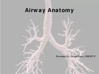
Slide14Artificial airway management
Airway management
indicated
in patients with loss of consciousness, facial or oral trauma, aspiration, tumor, infection, copious respiratory secretion, respiratory distress and the need for mechanical ventilation.
Slide15Types of airways
Oropharyngeal
airway
– curved plastic device inserted through the mouth and positioned in the posterior pharynx
Usually a short term use in the unconscious patient
Not used after oral surgery, or if loose teeth
Does not protect against aspiration
Slide16Slide17Nasopharyngeal airway
(nasal trumpet) – soft rubber or plastic tube inserted through nose into posterior pharynx
Facilitates frequent nasopharyngeal suctioning
Select size that is slightly smaller than diameter of nostril and slightly longer than distance from tip of nose to earlobe
Check nasal mucosa for irritation or ulceration and clean airway with hydrogen peroxide and water.
Slide18laryngeal mask airway
Composed of a tube with a cuffed mask like projection at the distal end
Inserted through the mouth into the pharynx
Seals the larynx and leaves distal opening of tube just above glottis
Easier placement than ET tube because visualization of vocal cords is not necessary
May cause
laryngospasm
and bronchospasm
Slide19Slide20Combitube
double – lumen tube
The distal tube enters the esophagus, where the cuff is inflated and ventilation is provided through the proximal tube, which opens at the level of the larynx
In rare instance where the distal tube
intubates
the trachea, ventilation is provided through the distal tube while the proximal tube is clamped.
Slide21Slide22Slide23Slide24ENDOTRACHEAL TUBE
Flexible tube inserted through the mouth and into the trachea beyond the vocal cords that acts as an artificial airway
Maintains a patent airway
Allows for deep tracheal suction and removal of secretions
Permits mechanical ventilation
Inflated balloon seals of trachea so aspiration from the GI tract cannot occur
Generally easy to insert in an emergency, but maintaining placement is more difficult so this is not for long term use
Slide25Slide26Surgical interventions
Tracheostomy
Airway
stenting
Slide27A Tracheostomy is a surgical opening in the anterior wall of the trachea to facilitate breathing. The tube enables airflow to enter the trachea and lungs directly, thus bypassing the pharynx and larynx.
Slide28Slide29Surgical techniques for the insertion of
tracheostomy
tubes
There are differing surgical techniques for the insertion of Tracheostomy tubes
Cricothyroidotomy
-
Cricothyroidotomy
-is an incision made through the skin and
cricothyroid
membrane to establish a patent airway during certain life-threatening situations
Percutaneous
tracheostomy
Tracheostomy
Slide30Slide31Slide32Percutaneous
tracheostomy
Percutaneous
Tracheostomy is an alternative to surgical Tracheostomy and is performed using a guide wire and a process of gradual dilation of the trachea and surrounding tissue. A
tracheostomy
tube is then inserted between the first and second or the second and third tracheal rings.
Slide33Slide34Complication of Tracheostomy
Immediate (Post Insertion)
Haemorrhage
(minor or severe)
Surgical emphysema
Delayed (Post Insertion)
Tube blockage with secretions. May be sudden or gradual
Infection of the stoma site and bronchial tree
Tracheal ulceration and Tracheal necrosis
Tracheo-oesophageal
fistula formation
Accidental
decannulation
,
Haemorrhage
(minor or severe)
Late (Post
Decannualtion
)
Tracheal dilation
Tracheal
stenosis
at the cuff site
Scar formation
Tracheomalacia
Slide35Slide36Tracheostomy tube
Firm, curved artificial airway inserted directly into the trachea at the level of the second or third tracheal ring through surgically made incision
Permits mechanical ventilation and facilitates secretion removal
Can be for long term use
Bypasses upper airway defenses, increasing susceptibility to infection
Allows the patient to eat and swallow
Slide37Tracheostomy tubes consist of three parts
An outer
cannula
with flange (neck plate) – the outer
cannula
is the outer tube that holds the
tracheostomy
open. A neck plate extends from the sides of the outer tube and has holes to attach cloth ties or strap around the neckInner cannula
– it fits inside the outer
cannula
. It has a lock to keep it from being coughed out, and it is removed for cleaning
An
obturator
– is use do insert a
tracheostomy
tube.
Slide38Types of Tracheostomy Tubes
Single Lumen Tubes
Double Lumen Tubes (inner
cannula
)
Uncuffed
Tubes or Cuffed Tubes
Fenestrated Tubes
Metal
tracheostomy
tubes
Slide39Cuffed tubes
It is having cuff ,which is located on the lower part of outer
cannula
which seals and gives ventilation.
Ventilator and other respiratory supportive devices can be attach to it
The cuff blocks any air from flowing around the tube and assures that the patient is well oxygenated.
All the air must therefore flow in and out through the tube itself. A pilot tube attached to the cuff stays outside the body and is used to inflate or deflate the cuff.
Slide40Slide41Fenestrated tube
A fenestrated tube has an opening (fenestration) in the back of the outer
cannula
.
The front of the tube can be blocked which allows the air to flow upwards to the upper part of the trachea and larynx.
A fenestrated tube allows the patient to breathe normally and speak or cough through the mouth.
A fenestrated
trach
tube is often used as the final step before
trach
tube removal.
It permits the patient to speak and cough on their own, providing an experimental trial for life after the
trach
tube.
Slide42Slide43Cuffless
tubes
Cuffless
tubes are primarily used in non-ventilated patients that have no difficulty swallowing and have no danger of aspiration.
Since there is no cuff, it allows air to pass into the upper trachea and larynx so the patient can cough and speak normally.
Cuffless
tubes are usually worn over a long period of time so require a very accurate fit in order to prevent pressure sores in the trachea or at the tracheal stoma.
Slide44Slide45Single lumen and double lumen
tracheostomy
tubes
Slide46Metal
tracheostomy
tube
Indication
Recommendation
Not used as frequently anymore.
Patients cannot get an MRI.
One needs to notify the security personnel at the airport prior to metal detection screening.
Slide47Pressure monitoring
Slide48The Passy-Muir Valve and open valve
Invented by a patient named David Muir, the Passy-Muir
Tracheostomy & Ventilator Swallowing and Speaking Valve is a simple medical device used by
tracheostomy
and ventilator patients.
The cuff has to be deflated while using valve
When placed on the hub of the
tracheostomy
tube the Passy-Muir Valve redirects air flow through the vocal folds, mouth and nose enabling voice and improved communication.
Slide49Weaning from
tracheostomy
If the patient can adequately exchange air and expectorate secretions, the
tracheosomy
tube can be removed
The stoma is covered with an occlusive dressing
The dressing must be changed if soiled or wetInstruct the patient to splint the stoma with fingers while speaking, swallowing or speaking
The opening will close in several days
Surgical intervention to close the opening is not required
Slide50Nursing diagnosis
Ineffective airway clearance related to presence of
tracheostomy
tube and difficulty expectorating sputum
Interventions
Auscultate
breath sounds
Remove secretions by suctioning to clear airwayEncourage slow deep breathing, turning and coughing to assist in mobilizing secretions
Position to alleviate
dyspnea
– head of the bed elevated 30-40 degree
Provide 100 % humidified oxygen
Slide51Ineffective therapeutic regimen management related to lack of knowledge about care of
tracheostomy
at home
Interventions
Demonstrate skill for the patient
Give clear, step by step instructions
Provide written information
Provide practice sessions
Explain regarding disease condition and the management done – so the patient will get an overall idea for self care
Instruct the patient to watch signs and symptoms and secretions
Slide52Impaired verbal communication related to use of cuffed artificial airway
Interventions
Listen attentively
Use picture board
Provide information to patient about condition
Provide reassurance about patients condition to relive fear and frustration
Provide information to patient regarding different types of
tracheostomy
tubes and speaking valve.
Slide53Risk for infection related to bypass of upper airway defense mechanisms and impaired skin integrity
Interventions
Monitor for systemic and localized signs and symptoms of infection
Monitor complete blood count
Maintain sterile technique when suctioning and providing Tracheostomy care
Provide trachea care every 4 – 8 hours as appropriate – clean the inner
cannula
, clean and dry the area around the stoma and change
tracheostomy
ties.
Inspect the area around the tube insertion site for redness and skin breakdown
Slide54Impaired swallowing related to
tracheostomy
tube
Interventions
Determine patients ability focus attention on learning/performing eating and swallowing tasks
Deflate the cuff before swallowing
Close the stoma site during swallowing
Monitor body weight to determine need for enteral feedings to maintain nutrition