
Foucault C Brouqui P Raoult D Bartonella quintana Characteristics and Clinical Management Emerg Infect Dis 2006122217223 httpsdoiorg103201eid1202050874 ID: 1038925
Download Presentation The PPT/PDF document "Figure 4 Figure 4. Western blot..." is the property of its rightful owner. Permission is granted to download and print the materials on this web site for personal, non-commercial use only, and to display it on your personal computer provided you do not modify the materials and that you retain all copyright notices contained in the materials. By downloading content from our website, you accept the terms of this agreement.


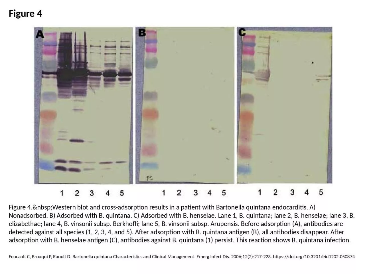
1. Figure 4Figure 4. Western blot and cross-adsorption results in a patient with Bartonella quintana endocarditis. A) Nonadsorbed. B) Adsorbed with B. quintana. C) Adsorbed with B. henselae. Lane 1, B. quintana; lane 2, B. henselae; lane 3, B. elizabethae; lane 4, B. vinsonii subsp. Berkhoffi; lane 5, B. vinsonii subsp. Arupensis. Before adsorption (A), antibodies are detected against all species (1, 2, 3, 4, and 5). After adsorption with B. quintana antigen (B), all antibodies disappear. After adsorption with B. henselae antigen (C), antibodies against B. quintana (1) persist. This reaction shows B. quintana infection.Foucault C, Brouqui P, Raoult D. Bartonella quintana Characteristics and Clinical Management. Emerg Infect Dis. 2006;12(2):217-223. https://doi.org/10.3201/eid1202.050874