
JEREMY E RADUCHA MD BRIAN COHEN MD TRAVIS BLOOD MD JULIA KATARINCIC MD ABSTRACT Brachial plexus injuries during the birthing process can leave infants with upper extremity de31cits corres ID: 938141
Download Pdf The PPT/PDF document "A Review of Brachial Plexus Birth Palsy ..." is the property of its rightful owner. Permission is granted to download and print the materials on this web site for personal, non-commercial use only, and to display it on your personal computer provided you do not modify the materials and that you retain all copyright notices contained in the materials. By downloading content from our website, you accept the terms of this agreement.

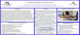
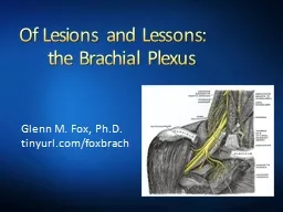
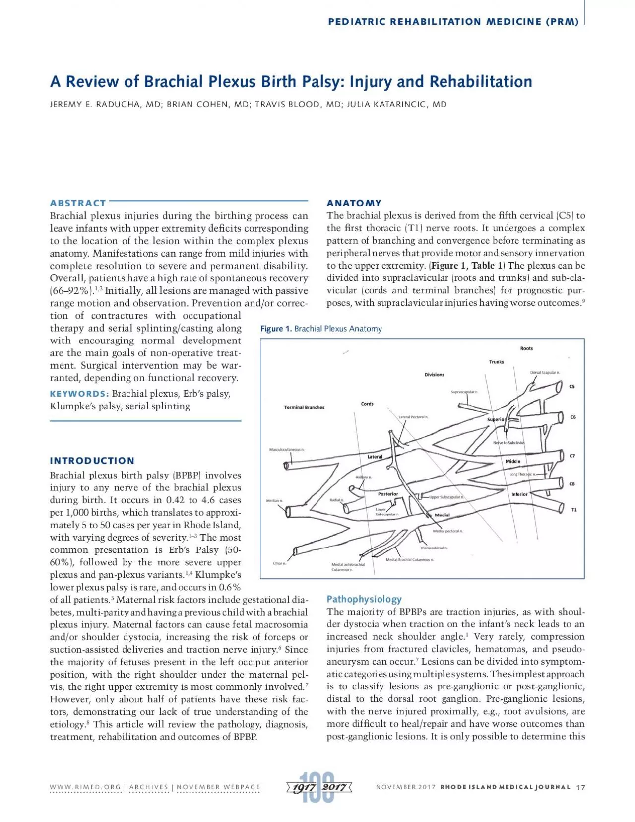
A Review of Brachial Plexus Birth Palsy: Injury and Rehabilitation JEREMY E. RADUCHA, MD; BRIAN COHEN, MD; TRAVIS BLOOD, MD; JULIA KATARINCIC, MD ABSTRACT Brachial plexus injuries during the birthing process can leave infants with upper extremity de cits corresponding to the location of the lesion within the complex plexus anatomy. Manifestations can range from mild injuries with complete resolution to severe and permanent disability. Overall, patients have a high rate of spontaneous recovery (66–92%). 1,2 range motion and observation. Prevention and/or correc - tion of contractures with occupational therapy and serial splinting/casting along with encouraging normal development are the main goals of non-operative treat - ment. Surgical intervention may be war- ranted, depending on functional recovery. KEYWORDS: Brachial plexus, Erb’s palsy, Klumpke’s palsy, serial splinting INTRODUCTION injury to any nerve of the brachial plexus during birth. It occurs in 0.42 to 4.6 cases per 1,000 births, which translates to approxi - mately 5 to 50 cases per year in Rhode Island, with varying degrees of severity. 1–3 The most common presentation is Erb’s Palsy (50- 60%), followed by the more severe upper plexus and pan-plexus variants. 1,4 Klumpke’s lower plexus palsy is rare, and occurs in 0.6% of all patients. 5 Maternal risk factors include gestational dia- betes, multi-parity and having a previous child with a brachial plexus injury. Maternal factors can cause fetal macrosomia and/or shoulder dystocia, increasing the risk of forceps or suction-assisted deliveries and traction nerve injury. 6 Since the majority of fetuses present in the left occiput anterior position, with the right shoulder under the maternal pel - vis, the right upper extremity is most commonly involved. 7 - tors, demonstrating our lack of true understanding of the etiology. 8 This article will review the pathology, diagnosis, treatment, rehabilitation and outcomes of BPBP. ANATOMY The brachial plexus is derived from the fth cervical (C5) to the rst thoracic (T1) nerve roots. It undergoes a complex pattern of branching and convergence before terminating as peripheral nerves that provide motor and sensory innervation to the upper extremity. ( Figure 1, Table 1 ) The plexus can be divided into supraclavicular (roots and trunks) and sub-cla - vicular (cords and terminal branches) for prognostic pur - poses, with supraclavicular injuries having worse outcomes. 9 Figure 1. Brachial Plexus Anatomy Pathophysiology The majority of BPBPs are traction injuries, as with shoul - der dystocia when traction on the infant’s neck leads to an increased neck shoulder angle. 1 Very rarely, compression injuries from fractured clavicles, hematomas, and pseudo- aneurysm can occur. 7 Lesions can be divided into symptom - atic categories using multiple systems. The simplest approach is to classify lesions as pre-ganglionic or post-ganglionic, distal to the dorsal root ganglion. Pre-ganglionic lesions, with the nerve injured proximally, e.g., root avulsions, are more dif cult to heal/repair and have worse outcomes than NOVEMBER 2017 RHODE ISLAND MEDICAL JOURNAL PEDIATRIC REHABILITATION MEDICINE (PRM) WWW.RIMED.ORG | ARCHIVES | NOVEMBER WEBPAGE 17 classi cation after advanced imaging. The Sunderland clas - si cation ( Table 2 ) categorizes nerve injuries based on the nerve structures damaged, ranging in severity from neuro - praxia to neurotmesis. 10 As expected, patients with less severe damage, e.g., neuropraxia, have a better chance at recovery. The most common way to describe BPBPs is based on the nerve roots involved, which can be detected by phys - ical examination. Upper trunk (Erb-Duchenne) palsies involve only the disruption of input from the C5 and C6 nerve roots. Upper plexus palsies involve roots C5, C6 and C7, with the addition of more distal de cits. Lower plexus (Klumpke’s) palsies involve the C8 and T1 nerve roots and can also affect the sympathetic chain with pre-ganglionic injuries. The most severe is the all-encompassing pan-plexus injury involving nerve roots C5-T1, with disruption to all functions of the upper extremity. Table 1. Brachial Plexus Functions Branching Location NerveRootInnvervationMuscle action Roots Dorsal Scapular n.
M: Rhomboid mm. and Levator Scapulae m. Rhom: scapular retraction, Levator=scapular elevation Long Thoracic n.C5,C6 & C7M: Serratus anterior m.Scapular protraction First intercostal n.T1M: intercostal m. n/a Trunks Suprascapular n. C5, C6 M: Supraspinatus m, Infraspinatus m. S: Shoulder joint capsule Supra= Arm abduction. Infra= Arm external rotation Nerve to SubclaviusC5, C6M: Subclavius m. n/a Divisions none Cords Posterior Upper Subscapular nC5-T1Motor: Upper subscapularis m. Arm internal rotation Lower Subscapular nC5-T1 Motor: Lower Subscapularis m., Teres Major m. LS= Arm internal rotation Thoracodorsal n. C5-T1Motor: Latissimus dorsi m. Arm adduction LateralLateral Pectoral n.C5-C7Motor: Pectoralis Major m. Arm Adduction MedialMedial Pectoral n. C8-T1 Motor: Pectoralis Major m., Pectoralis Minor m. Arm Addution Medial Brachial cutaneous n. C8-T1Sensory: medial armn/a Medial Antebrachial cutaneous n. C8-T1Sensory: medial forearmn/a Terminal Nerves Radial n. C5-T1 Motor: Triceps mm, brachioradialis m., ECRL, ECRB, ECU, EDC, EIP, EDM, EPL, EPB, APL, Supinator m., Finger extensors Sensory: posterior brachial cutaneous, inferior lateral brachial cutaneous, posterior antebrachial cutaneous, superficial radial (post. radial hand) Elbow extension, Wrist extension, Finger Extension, Thumb extension, thumb abduction, Forearm supination; Brachioradialis=elbow flexion Axillary n. C5-T1 Motor: Deltoid m., Teres Minor m., Sensory: Lateral proximal arm Delt= Arm abduction, Teres= Arm external rotation Musculocutaneous n. C5-C7 Motor: Biceps brachii m., Brachialis m, corocobrachialis m. Sensory: Lateral cutaneous n. of the forearm Elbow Flexion, Forearm supination S: lateral forearm Median n. C5-T1 Motor: FCR, Palmaris longus m., FDS, radial 1/2 FDP, Pronator teres m. FPL, Pronator quadratus m., FPB (superficial head), Opponens pollicis, APB, 1st-2nd lumbricals Sensory: Radial 3 1/2 fingers, palmar cutaneous branch Wrist flexion, Forearm pronation, thumb flexion/abduction/opposition, finger PIP flexion, IF/MF MCP and DIP flexion Ulnar n. C8-T1 Motor: FCU, ulnar 1/2 FDP, Flexor DM, Abductor DM, Opponens DM, Adductor pollicis, FPB (deep head), Palmaris brevis m. Dorsal interossei mm. Palmar interossei mm., 3rd-4th Lumbricals Sensory: Dorsal ulnar cutaneous n., Palmar ulnar cutaneous n. Wrist flexion, Thumb adduction/flexion, SF flexion/abduction/opposition, finger adduction/abduction, 4th and 5th finger DIP/MCP flexion Type of Nerve Injury Prognosis Neuropraxia Stretch injury with intact nerve continuity Spontaneous recovery likely Axonotmesis Axonal injury with intact nerve sheath Variable recovery Neurotmesis Complete nerve rupture; neither axon nor sheath intact Poor prognosis for spontaneous recovery Table 2. Sunderland Classi cation Sunderland SS. The anatomy and physiology of nerve injury. Muscle Nerve . 1990;13(9):771-784. doi:10.1002/mus.880130903 PEDIATRIC REHABILITATION MEDICINE (PRM) WWW.RIMED.ORG | ARCHIVES | NOVEMBER WEBPAGE 18 DIAGNOSIS Maternal history, physical examination and diagnostic imag - ing can provide a wealth of information to make the proper diagnosis and injury classi cation. The patient’s mother should be interviewed for the BPBP risk factors mentioned previously. Abnormal primitive re exes, e.g., Moro re ex and tonic neck re ex, are often the rst clues in the new - born examination. It is also important to palpate the infant’s clavicle and humerus, as fractures can affect upper extremity movements and be confused with brachial plexus palsy. A septic shoulder and isolated radial nerve palsy should also be in the differential diagnosis, but they are less common and are associated with different physical and laboratory ndings. Depending where the lesion is located, the patient’s affected extremity will present in different positions. With Erb’s palsy (C5-6), the arm is adducted and internally rotated at the shoulder and extended at the elbow, due to weakness in the deltoid, supero-posterior rotator cuff and biceps. A patient with upper plexus palsy (C5-7) has the above pos - ture as well as wrist and ngers exion due to radial nerve involvement and wrist/ nger extensor weakness. Pan-plexus injuries (C5-T1) typically present with a accid extr
emity. Pre-ganglionic injuries, which carry a worse prognosis,, may lead to head tilting to the opposite side (denervation of paraspinal musculature), medial winging of the scapula, diaphragm dysfunction, and Horner’s syndrome. As children age, their disabilities become more apparent. Scoring systems such as the Toronto Test Score, Active Movement Scale, and Modi ed Mallet system have been developed to grade and track upper extremity function. 11 The Modi ed Mallet score is the most commonly used when evaluating older children ( 3 years old). It uses ve categories to assess shoulder function, with a 0–5 grading for each category. Higher scores correlate to higher function, but the examination requires patient participation and is heavily weighted toward shoulder external rotation. Imaging can help clarify the diagnosis and classi ca - tion. Initially, radiographs of the upper extremity should be obtained to rule out fractures, which could be confused with or occur concomitantly with brachial plexus palsy. MRI and CT myelography can be used to detect root avulsions. Elec - tromyography has been suggested if there is no nerve recov - ery by 6 months of age, in order to detect a pre-ganglionic injury, which is potentially amenable to operative interven - tion. Other evidence shows, however, that electromyography can be discordant from clinical bicep function at 3 months of age and the test may not be a reliable indicator for surgery. 1 TREATMENT AND OUTCOMES Treatment for a suspected brachial plexus palsy should begin immediately with frequent, passive range of motion of the affected upper extremity. Parents should be instructed to range both arms at every diaper change to make it a daily rou - tine and encourage compliance. Some authors recommend a two-week period of immobilization to promote healing and decrease pain, 9 but others nd little evidence that immobi - lization has any bene t. 1 Contractures can begin as early as 2-3 weeks after birth, with the glenohumeral joint most commonly affected. Without early treatment, the contrac - tures can progress rapidly and cause posterior subluxation/ dislocation of the humeral head. 1,9,12 After an initial observation period, children can be cate - gorized as having either partial or total paralysis. Patients with total paralysis should be referred to a tertiary center for early surgical evaluation, as they have a very low likelihood of spontaneous recovery. Patients with partial paralysis have a higher chance of recovery, and there is complete recovery by 3 months of age with as many as 92% of these patients. 9,13 Other evidence suggests complete recovery rates may not be as high as originally thought, with as many as 20–30% of patients having a long-term defecit. 1,2 Patients who do not have complete recovery by 1 month of age should be evaluated by a pediatric therapist for continued monitoring and rehabilitation. Many physicians use the lack of antigrav - ity biceps function return by 3 months as an indication for nerve surgery since it is a poor prognostic indicator for com - plete spontaneous recovery; 1,4 however, this does not pre - clude good functional recovery. Recovery of wrist extension is also a positive prognostic sign. Other physicians advocate continued rehabilitation until at least 6 months of age before considering surgery. Some evidence has shown that children who recovered antigravity biceps function between the 3 rd and 6th months of life always had an incomplete recovery compared to those who regained function prior to 3 months. 9 If children undergo surgery, it is typically performed between 3 and 8 months of age; earlier surgery (at 3 months) is indicated in children with pan-plexus palsies and Horner’s syndrome. The main goals of surgery, in order of importance, are to restore elbow exion, shoulder abduction, shoulder external rotation, wrist extension and hand function. 7 The options for early surgical intervention include direct nerve repair with resection and grafting, or nerve transfers from surrounding motor nerves. In spite of surgery, many patients still suffer some degree of long-term sequalae. 2 In patients who develop contracture or have persistent weakness, later surgery can be bene cial. Lysis of contractures, osteotomies and local tend
on transfers can help return functional motion and correct deformities. 12,14 Children who recover meaningful biceps function by 6 months of age are typically treated non-operatively with rehabilitation and monitoring. There is little high grade research discussing non-operative management techniques and protocols for brachial plexus birth palsies. All non-oper - ative treatment involves a multidisciplinary team approach, with occupational therapy and splinting to prevent or correct contractures. The goals of treatment prior to muscle func - tion recovery are to prevent contracture, strengthen recov - ering muscles, stimulate sensory nerves, and encourage the achievement of normal developmental milestones. As the PEDIATRIC REHABILITATION MEDICINE (PRM) WWW.RIMED.ORG | ARCHIVES | NOVEMBER WEBPAGE 19 child grows, passive range of motion should be transitioned to participation in age-appropriate activities for rehabilita - tion with regular follow-up to assess functional scores, arm growth and joint integrity. Elbow exion contractures are a fairly common occurrence, even with triceps sparing pal - sies. For children who develop contractures, stretching and serial night splinting can be used for contractures less than 20 degrees. Treating deformities in this range will prevent progression and help cosmetic appearance, as the elbow’s functional range of motion is between 30–130 degrees. 15 Serial casting and splinting of elbow exion contractures can yield good results, but this approach can be compli - cated by radial head dislocation, bony ingrowth at the joint, and loss of elbow exion while gaining extension. Much of the research is focused on elbow exion contractures but contractures preventing forearm supination and shoulder external rotation are also commonly present. A pilot study has shown improvement in Toronto and Active Movement Scales of supination and shoulder external rotation with a Supination-External rotation orthosis worn 22 hours per day with reprieves for therapy twice per day. 16 Botulinum toxin injections with serial casting have also shown promise in patients who failed serial casting alone. 17 The toxin relaxes the antagonist muscle at the contracted joint, particularly in cases of co-contraction, a common long-term complication. Despite the lack of consensus regarding surgical indica - tions and rehabilitation protocols, patients do have good long-term outcomes. Most studies show the majority of patients are independent in activities of daily living, even with persistent functional de cits. 1,2,7,8,13,19 In a subjective study of adolescents, all patients reported a ‘really good’ quality of life, but they were also all dissatis ed with their current condition and hoped for continued improvement. 20 CONCLUSION Brachial plexus birth palsies can be stressful and challeng - ing for parents and children. Despite a better understanding of the pathology and treatment options, injury incidence has remained unchanged. 1–3 In general, upper plexus palsies recover better than lower plexus and pan-plexus palsies; neuropraxia does better than neurotmesis; and post-gan - glionic lesions recover better than pre-ganglionic lesions. Care should continue to focus on early identi cation and therapy to minimize complications. Early referral to tertiary centers is crucial, as a multi-disciplinary approach can help promote recovery and prevent complication. Fortunately there is a high rate of spontaneous recovery, but for patients who don’t recover spontaneously there are non-surgical and surgical options to improve functional outcomes and pre - vent devastating contractures. With an ever-expanding body of research geared towards improving care and knowledge of the injury, the future should show improved long-term outcomes for these patients. References 1. Malessy MJA, Pondaag W. Obstetric Brachial Plexus Inju - ries. Neurosurg Clin N Am . 2009;20(1):1-14. doi:10.1016/j. nec.2008.07.024. 2. Hoeksma AF, Wolf H, Oei SL. Obstetrical brachial plexus inju - ries: incidence, natural course and shoulder contracture. Clin Rehabil . 2000;14(5):523-526. doi:10.1191/0269215500cr341oa. 3. Evans-Jones G, Kay SPJ, Weindling AM, et al. Congenital bra - chial palsy: incidence, causes, and outcome in the United Kingdom and Republic of
Ireland. Arch Dis Child Fetal Neo - natal Ed . 2003;88(3):F185-9. http://www.ncbi.nlm.nih.gov/ pubmed/12719390. Accessed May 15, 2017. 4. Kozin SH. Brachial Plexus Microsurgical Indications. J Pediatr Orthop . 2010;30:S49-S52. doi:10.1097/BPO.0b013e3181cd9ed2. 5. al-Qattan MM, Clarke HM, Curtis CG. Klumpke’s birth palsy. Does it really exist? J Hand Surg Br . 1995;20(1):19-23. http:// www.ncbi.nlm.nih.gov/pubmed/7759926. Accessed May 15, 2017. 6. Gilbert WM, Nesbitt TS, Danielsen B. Associated factors in 1611 cases of brachial plexus injury. Obstet Gynecol . 1999;93(4):536- 540. http://www.ncbi.nlm.nih.gov/pubmed/10214829. Accessed May 15, 2017. 7. Shenaq SM, Bullocks JM, Dhillon G, Lee RT, Laurent JP. Man - agement of infant brachial plexus injuries. Clin Plast Surg . 2005;32(1):79-98. doi:10.1016/j.cps.2004.09.001. 8. Hale HB, Bae DS, Waters PM. Current concepts in the man - agement of brachial plexus birth palsy. J Hand Surg Am . 2010;35(2):322-331. doi:10.1016/j.jhsa.2009.11.026. 9. Abid A. Brachial plexus birth palsy: Management during the rst year of life. Orthop Traumatol Surg Res . 2016;102(1 Sup - pl):S125-32. doi:10.1016/j.otsr.2015.05.008. 10. Sunderland SS. The anatomy and physiology of nerve injury. Muscle Nerve . 1990;13(9):771-784. doi:10.1002/mus.880130903. 11. Duff S, DeMatteo C. Clinical Assessment of the Infant and Child Following Perinatal Brachial Plexus Injury. J Hand Ther . 2015;28(2):126-134. doi:10.1016/j.jht.2015.01.001. 12. Abid A, Accadbled F, Louis D, et al. Arthroscopic release for shoulder internal rotation contracture secondary to brachi - al plexus birth palsy: clinical and magnetic resonance imag - ing results on glenohumeral dysplasia. J Pediatr Orthop B . 2012;21(4):305-309. doi:10.1097/BPB.0b013e328353688e. 13. Laurent JP, Lee R, Shenaq S, Parke JT, Solis IS, Kowalik L. Neu - rosurgical correction of upper brachial plexus birth injuries. J Neurosurg . 1993;79(2):197-203. doi:10.3171/jns.1993.79.2.0197. 14. Abzug JM, Kozin SH. Evaluation and Management of Brachial Plexus Birth Palsy. Orthop Clin North Am . 2014;45(2):225-232. doi:10.1016/j.ocl.2013.12.004. 15. Ho ES, Roy T, Stephens D, Clarke HM. Serial casting and splint - ing of elbow contractures in children with obstetric brachial plexus palsy. J Hand Surg Am . 2010;35(1):84-91. doi:10.1016/j. jhsa.2009.09.014. 16. Durlacher KM, Bellows D, Verchere C. Sup-ER orthosis: an in - novative treatment for infants with birth related brachial plexus injury. J Hand Ther . 2014;27(4):335-39; quiz 340. doi:10.1016/j. jht.2014.06.001. 17. Basciani M, Intiso D. Botulinum toxin type-A and plaster cast treatment in children with upper brachial plexus palsy. Pediatr Rehabil . 2006;9(2):165-170. doi:10.1080/13693780500402229. 18. Kerr C, McDowell B, Cosgrove A, Walsh D, Bradbury I, Mc - Donough S. Electrical stimulation in cerebral palsy: a random - ized controlled trial. Dev Med Child Neurol . 2006;48(11):870- 876. doi:10.1017/S0012162206001915. 19. DiTaranto P, Campagna L, Price AE, Grossman JAI. Outcome following nonoperative treatment of brachial plexus birth inju - ries. J Child Neurol . 2004;19(2):87-90. doi:10.1177/0883073804 0190020101. 20. Squitieri L, Larson BP, Chang KWC, Yang LJS, Chung KC. Un - derstanding quality of life and patient expectations among ado - lescents with neonatal brachial plexus palsy: a qualitative and quantitative pilot study. J Hand Surg Am . 2013;38(12):2387- 2397.e2. http://www.ncbi.nlm.nih.gov/pubmed/24416766. Ac - cessed May 8, 2017. PEDIATRIC REHABILITATION MEDICINE (PRM) WWW.RIMED.ORG | ARCHIVES | NOVEMBER WEBPAGE 20 Authors Jeremy E. Raducha, MD; Department of Orthopaedic Surgery, Warren Alpert Medical School of Brown University, Providence, RI. Brian Cohen, MD; Department of Orthopaedic Surgery, Warren Alpert Medical School of Brown University, Providence, RI. Travis Blood, MD; Department of Orthopaedic Surgery, Warren Alpert Medical School of Brown University, Providence, RI. Julia Katarincic, MD; Department of Orthopaedic Surgery, Warren Alpert Medical School of Brown University, Providence, RI. Correspondence Jeremy E. Raducha, MD Department of Orthopaedic Surgery Rhode Island Hospital 593 Eddy Street Providence, RI 02903 401-444-4030 Fax 401-444-6182 Jeremy.raducha@gmail.com PEDIATRIC REHABILITATION MEDICINE (PRM) WWW.RIMED.ORG | ARCHIVES | NOVEMBER WEBPAGE 21