
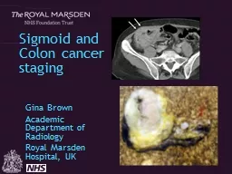
Gina Brown Academic Department of Radiology Royal Marsden Hospital UK Dukes Histological system for rectal cancers extrapolated for colon cancers 5 year survival 81 if confined to bowel wall ID: 908209
Download Presentation The PPT/PDF document "Sigmoid and Colon cancer staging" is the property of its rightful owner. Permission is granted to download and print the materials on this web site for personal, non-commercial use only, and to display it on your personal computer provided you do not modify the materials and that you retain all copyright notices contained in the materials. By downloading content from our website, you accept the terms of this agreement.


Slide1
Sigmoid and Colon cancer staging
Gina BrownAcademic Department of RadiologyRoyal Marsden Hospital, UK
Slide2Dukes Histological system for rectal cancers extrapolated for colon cancers5 year survival:
81% if confined to bowel wall64% if invasion through the wall27% if local lymph nodes involvedAJCC TNM staging systemT stage, N stage, M stage7th Edition [Edge and Compton, 2010]
Staging of colon cancers
Slide3Extramural Vascular Invasion (EMVI)
Reduced 5 year survival Depth of extramural spreadHermanek divided T3 tumours into 4 groupsInvolvement of Non Peritonealised Resection Margin Very high risk local recurrenceHistological gradeWell differentiated, 76% 5 year survival
Poorly differentiated, 31% 5 year survivalOther prognostic factors
Slide4How often are prognostic factors reported preoperatively in colon cancer?
- EMVI- depth of extramural spread in mm - non-peritonealised resection margin- transperitoneal
breach?
Slide5Currently: no role for imaging for local staging of colon cancers?
Slide6Survival
Colon Cancer
Age-Standardised Five-Year Relative Survival Rates
England and Wales 1971-1995, England 1996-2009
Rectal Cancer
Age-Standardised Five-Year Relative Survival Rates
England and Wales 1971-1995, England 1996-2009
Cancer Research UK
Slide7MRI based
Selection
of patientsFor range treatments
Local excision
MRI and PET surveillance
Deferral of surgery
Chemoradiotherapy
Restage:
Timing of
surgery
after CRT
6 vs 12?
Biological agents and neoadjuvant
chemotherapy for MRI EMVI
Further Therapy
/Extended surgery
for mrCRM/low rectal
MRI T1/T2 Nx
EMS /TEMS
pre/post operative CRT
MRI surveillance…
MRI Low rectal
Stage 3 or 4
Post CRT
yMRI TRG 1-2
MRI T3a/T3b N any
Low rectal stage 1/2
Primary TME Surgery: open v laparoscopic
MRI T3c/T3d N any
EMVI positive CRM safe
potential CRM unsafe
Treatment options for
Rectal Cancer
Palliative Chemotherapy
Metastatectomy
Primary colon resection:
laparoscopic/open
CT Staging
Metastatic
disease?
Yes/No
80-90%
10-20%
Treatment options for
Colon Cancer
Slide8Colon Cancer has a high recurrence rate.
O’Connell 2008 ACCENT Data Setn=17,381recurrence= 5,722 (32%)
J Clin Oncol. 2008 May 10;26(14):2336-41.
Slide9Metaanalysis
Slide10Slide11Nodal Staging
Slide12Meta-analysis conducted on studies assessing accuracy of CT in staging colorectal cancer to detect tumour invasion beyond MP :
Sensitivity is as high as 86%.Specificity of 78%The ability of CT to predict the nodal status is however poor.However none of the studies ever looked at the ability of CT to predict prognosis.
Dighe S, Purkayastha S, Swift I, Tekkis PP, Darzi A, A'Hern R, Brown G: Diagnostic precision of CT in local staging of colon cancers: A Meta analysis.
Clin Radiol. 2010 Sep;65(9):708-19.
Slide13Good prognosis T2/early T3
Slide14T3 good tumour
Slide15Understanding T4 disease
Slide16Slide17Poor prognosis
Slide18*
Slide19Poor prognosis
Slide20CT staging of colons
To examine whether the radiological features of the primary colonic tumour seen on the pre-operative CT scan could be used to predict clinical outcome.To compare pre-operative CT-based prognostication with post-operative histology
Smith N, Bees, N. Predicting Prognosis in Colon Cancer: Validation of a New Preoperative CT Staging Classification and Implications for Clinical Trials. Colorectal Disease 2006;
8
Slide21126 scans analysed
Slide22Prognostic score
Histological variable
Good prognosis
Poor prognosis
T stage
T1, T2 or T3<5mm
T3>5mm or T4
N stage
N0, N1
N2
EMVI
Absent
Present
Slide23Identification of poor prognosis tumours
56% (70/126) had CT defined poor prognosis tumours
Slide24T staging / prognosis
Stage-for-stage accuracy=60.3%Poor prognosis (Stage T3/T4, N2, EMVI)Overall Accuracy=83.3% (Sensitivity=92.4%; Specificity=42.1%)Positive Predictive Value=89.8%; Negative Predictive Value=50.0%
Slide25Slide26CT prediction of prognosis
Slide27the depth of tumour invasion beyond the muscularis propria (MP) as seen on CT and demonstrated excellent correlation with histology.
T1/T2 + T3 <5mm tumour invasion beyond MP (87% 3-year survival).T4+T3≥5mm tumour invasion beyond MP (53% 3 year survival).
Smith N, Bees, N. Predicting Prognosis in Colon Cancer: Validation of a New Preoperative CT Staging Classification and Implications for Clinical Trials.
Colorectal Disease
2006;
8
Slide28Can we refine the radiological definition of poor prognosis?
Involvement of peritoneal surfaces
Slide29Can we refine the radiological definition of poor prognosis?
Sensitivity: 78% Specificity: 67%
Accuracy: 74%
PPV: 81%
Dighe S, Blake H,
Koh
MD, Swift I,
Arnaout
A, Temple L,
Barbachano
Y, Brown G: Accuracy of
multidetector
computed tomography in identifying poor prognostic factors in colonic cancer.
Br J Surg. 2010 Sep;97(9):1407-15.
Slide30Can we refine the radiological definition of poor prognosis?
Involvement of the peritoneal and mesenteric surfacesLymph node involvementSensitivity 58%Specificity 64%
Slide31Can we refine the radiological definition of poor prognosis?
Data collectionInvolvement of the peritoneal and mesenteric surfacesLymph node involvement
Extramural venous invasion
Slide32Detection of EMVI using MDCT: high positive predictive value
Slide33Value of >5mm Extramural Depth of Spread using CT
77 % of patients (42 of 54)with a histologically poor prognosis were identified based on T category also 74 % of node-positive patients (29 of 39) compared with 58% by using size
Slide34FOxTROT trial design
3 Fu Ox
±
Pan
(6 weeks)
9 Fu Ox
(18 weeks)
12 Fu Ox (24 weeks)
±
Panitumumab (6 weeks)
CT staging
T3+ or N2+ colon cancer,
potentially curative
n=350
n=700
Primary outcome – freedom from disease at 2 years
R
a
n
d
o
m
is
e
S
u
r
g
S
u
r
g
Slide35End points of Foxtrot trial
1050 patients over 3 years (150 pilot + 900)for recurrence free survival; 80% power at p<0.05 to detect 25% proportional reduction in treatment failure, e.g. Recurrence reduced from 32% to 24%.
for tumour shrinkage; 90% power at p<0.01 to detect a small/moderate (0.3sd) difference in pathological tumour shrinkage with addition of panitumumab, i.e. Depth of invasion.
Slide36Imaging– what’s new in this trial?
New staging systemKnowledge and visualisation of peritoneal anatomyIdentification of poor prognostic features in vivoQuality assurance: workshops, detailed imaging data collection
Slide37This trial is thus reliant on the ability of the radiologists to identify a cohort of high risk patients suitable for randomisation to receive neoadjuvant therapy.
Slide38Summary colon cancer staging
Tumour morphology: annular, semi-annular, mucinous, ulceratingSite : caecum
, ascending, hep flexure, transverse, splenic flexure, descending, sigmoidBorder of infiltration: mesenteric
vs
peritonealised
Diameter and thickness
T
substage
(good or poor): T3<5mm or >5mm
Nodal and venous spread:
ileocolic
, middle colic, left colic,
sigmoidal
veins
Adjacent organ infiltration/perforation/obstructionSynchronous metastatic disease
Slide39Was CT successful in identifying high risk? Control arm pathology
49/50 – pT3/4 (98%)2643/50 – AJCC pTNM stage II/III high risk (86%)/50 –pNode positive (52%)10/50 – 20% pCRM
positive24/48 – (50%) pEMVI positive
Slide40Sigmoid Cancer is a problem
Dis Colon Rectum. 2010 Jan;53(1):57-64.
Slide41Recurrence sigmoid cancer
N=
Follow-up
Local recurrence colon
Local recurrence sigmoid
Cass
1976
Retrospective 1968-1974
280
Min 1 yr
22,5%
25%
Willett 1984
Retrospective
533
19%
21%
Sjövall 2007
Prospective 1996-2000
1,856
Min 3 yrs
11,5%
11,6%
Slide42MDT 2007-09
296 sigmoid cancers 104 for palliative careCurable sigmoid cancers: n=192No FU data at all: n=42With FU: n=150
FU 36 months (range 1-76, median 38)Recurrence: 62/192 (32%)
Local recurrence: 19 (11%)
Recurrence sigmoid cancer
Slide43High risk features
Tumour involving non peritonealised fascial marginTumour penetration of adjacent organs4 or more involved nodesExtramural venous invasionDepth of extramural spread >5mm
Slide44Burton 2006
Int. J. Radiation Oncology Biol. Phys
Slide45Primary surgery
n=5716 at/above peritoneal reflection19 rectosigmoid22 sigmoid
Neoadj CRTx + surgery n=18
9 at/above peritoneal reflection
5 rectosigmoid
4 sigmoid
Burton 2006
Int. J. Radiation Oncology Biol. Phys
Slide46MRI predicted prognosis with final histological prognosis in 57 patients undergoing primary surgery
84% (CI =72.6-92.7%) accuracy for MRI prediction of prognosis
Kappa = 0.63
Sensitivity = 90%
Specificity = 72%
Positive predictive value = 88%
Negative predictive value = 76%
Burton 2006
Int. J. Radiation Oncology Biol. Phys
Slide47Neoadjuvant Treatment
Burton 2006
Int. J. Radiation Oncology Biol. Phys
Slide48Pelvic sigmoid
Slide49Staging and treatment
Sigmoid colon has traditionally been grouped with the remainder of the colon Direct continuation of the rectum located in the pelvis treating sigmoid cancer Subject to the same constraints as rectal cancer with similar potential surgical challenges and risks of a threatened margin
Improved image quality in rectal has enabled better tumour depiction and superior risk stratification
Precise
imaging staging enables appropriate
surgical and oncological treatment planning
This could translate into a reduction in pelvic recurrence rates
Slide50Preoperative staging
Currently CT is widely used to assess sigmoid cancers, CT has limited ability to delineate pelvic structures and detailed anatomyHigh resolution MRI better suited evaluating pelvic structuresMay help to identify those at risk of incomplete resection/ local recurrenceSuch patients may benefit from radical neoadjuvant
treatment and more accurate surgical ‘road-mapping’
Slide51IMPRESS Trial
Hypothesis: Accurate preoperative imaging (MRI) will improve recurrence rate and survival through:
better surgical decision making
Greater proportion receiving radical treatment (
neoadjuvant
therapy or extended surgery)
Slide52Biopsy proven sigmoid cancer
OBSERVATIONAL PATHWAY
MRI is local policy
MDT review CT & MRI
INTERVENTIONAL PATHWAY
Randomised
to have MRI
Randomised
not to have MRI
MDT review CT & MRI
MDT review CT only
Treatment Outcomes
Slide53Endpoints IMPRESS Trial
Primary: Observational: Measure difference in detection of high risk patients between CT and MRI and the resultant difference in Rx strategy
Randomised: Compare the proportion of patients undergoing radical treatment in the two armsSecondary:
R
ecurrence
rate
at 1, 3 and 5 years
OS
and
DFS at 1, 3 and 5 years
A
ccuracy
of CT and MRI to identify poor prognosis
tumours
compared to the gold standard of histopathology Quality of surgery CRM positivity rates on pathology
Permanent defunctioning stoma rates
Slide54Study design
Observational and randomised arms (1:1)Expected improvement of 20% in sensitivity of detection of high risk patients, 97 patients need to be randomised to each armDrop out rate 20%243 patients needed in randomisation arm
Folllow-up 5 years, outcomes reported at 1, 3, and 5 years
Slide55Biopsy proven sigmoid cancer
OBSERVATIONAL PATHWAY
MRI is local policy
MDT review CT & MRI
INTERVENTIONAL PATHWAY
Randomised
to have MRI
Randomised
not to have MRI
MDT review CT & MRI
MDT review CT only
Treatment Outcomes
Slide56Sites
Open: RMHCroydonSalisburyHarrogate
St Mark’s
Opening:
Portsmouth
Taunton
Yeovil
Macclesfield
Scunthorpe
Manchester Royal Infirmary
Hinchingbrooke
East Kent
Leigton
North Tees
Royal Free
Slide57IMPRESS TrialIMProving
Radical treatment through MRI Evaluation of pelvic Sigmoid cancerS Contact
Gina Brown (Principal Investigator) Gina.Brown@rmh.nhs.uk
Lisa
Scerri
(Clinical Trial Coordinator)
lisa.scerri@rmh.nhs.uk
0208 915 6067
Slide58Sigmoid cancer
Sigmoid cancer has a high recurrence rateSigmoid cancer has a worse outcome than rectal cancerMRI is able to identify poor prognostic tumours preoperativelyPreoperative staging enhances optimal treatment strategy including neoadjuvant treatment
Sigmoid cancer with poor prognostic features should be discussed for neoadjuvant treatment (IMPRESS Trial)
Slide59Better staging Colon cancer: new treatment possibilities
MRI based
Selectionof patientsFor range treatments
MRI and PET surveillance
Screen for metastatic disease
Chemoradiotherapy
Restage:
Biological agents and neoadjuvant
chemotherapy for MRI EMVI
Further Therapy
/Extended surgery
MRI T1/T2/early T3
Primary Surgery: laparoscopic
MRI T3c/T3d N any
EMVI positive
CRM safe
MRI potential resection
margin
unsafe in rectosigmoid
MRI potential resection
margin
unsafe in colon
Extended surgery
Slide60Acknowledgements
Shwetal Dighe, Sarah Burton and Neil Smith, Chris Hunter, Ian Swift and Muti Abulafi and the Royal Marsden Hospital Colorectal Multidisciplinary NetworkFoxTrot trial co-investigators: D Morton, P Quirke, M Seymour, R Gray, L Magill.