
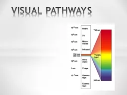
Perception of shape motion color Two pathways retina cortex visual perception retina brainstem diencephalon eye movements circadian ID: 784764
Download The PPT/PDF document "VISUAL PATHWAYS VISUAL SYSTEM" is the property of its rightful owner. Permission is granted to download and print the materials on this web site for personal, non-commercial use only, and to display it on your personal computer provided you do not modify the materials and that you retain all copyright notices contained in the materials. By downloading content from our website, you accept the terms of this agreement.


Slide1
VISUAL PATHWAYS
Slide2VISUAL SYSTEM
Perception
of
shape
motion
color
Two
pathways
retina –
cortex
visual
perception
retina –
brainstem
,
diencephalon
eye
movements
circadian
photoentrainment
accommodation
pupillary
reflexes
Slide3Light
passes
through
the
cornea
,
aqueous
humor,
lens
, and
vitreous
body
to
form
an
image on
the
retina.
Slide4Macula
lutea + fovea
centralis
=
areas
of
the
highest visual acuity
Fundus
oculi
Slide5RETINA
10
layers
:
mainly
separated
by cell
bodies (
nuclear layers) and axons (plexiform layers)
Slide65
main
cell
types
:
photoreceptors
bipolar cells horizontal cells
amacrine cells ganglion cells
Photoreceptors
:
rods
and
cones
involved
in
transduction
converting
the
light
signal
into
a
nerve impulse
Slide7neurons with serial
(
vertical
)
connection
the main visual pathway photoreceptors
→ bipolar cells → ganglion cells
neurons
with
parallel
(
horizontal
)
connection
modulation
of
the
visual
information
by retina
horizontal
cells
amacrine
cells
Slide8Slide9Cones
(7
million
)
cluster
at
fovea
(macula lutea)
detect color in bright light = photopic vision
Rods (100 million
)
outside
the
fovea
sensitive to
shape
and
movement
=
scotopic
vision
Slide10CONES
3
different
types
with
three different photopigments: blue, green and red
Each type is maximally sensitive to the
wavelength
that
corresponds
to
the
specific
color
range
(
spectral
sensitivity)
Slide11GANGLION CELLS
P
cells
(80%)ganglion cells
that
monitor
cones
smaller, more numerousaxons
end on parvocellular laminae of LGNprovide information about fine detail and
colorM cells (10%)ganglion cells
that
monitor
rods
relatively
large
axons
end on
magnocellular
laminae
of
LGN
provide
information
about
a
general
form
of
an
object
,
motion
, and
shadows
in
dim
light
non-P non-M
cells
(10%)
projection
to
subcortical
nuclei
,
koniocellular
cells
of LGN
Slide12PRIMARY VISUAL PATHWAY
The
primary visual
pathway
connects
the retina with lateral
geniculate nucleus and primary visual cortex (
retinogeniculostriate pathway) It is responsible
for
detection
of
shape
,
movement
and
color
1
st
neuron (
photoreceptors
)
2
nd
neuron
(bipolar cells)
3rd neuron (ganglion cells)
LGN
Optic
chiasm
Primary
visual cortex
CN II
Optic
tract
Optic
radiation
Slide13Slide14LGN
is
composed
of 6
layers
Layers
1 and 2
contain larger neurons Layers
3 - 6 contain smaller neurons
LATERAL GENICULATE NUCLEUS (LGN)
Slide15Slide16Ipsilateral input
enters
layers
2,3 and 5
Contralateral
input
enters layers 1, 4 and 6
Slide17LGN
contains
the
topographic
representation of what
the retina “ sees”. This retinotopic map is sent to the cortex.
LGN modulates and regulates the flow of visual information to the primary visual cortex cortex can control efficiency of thalamic input
Slide18optic radiation
(
geniculocalcarine
fibres
) runs
under the temporal lobe to the occipital lobe
GENICULOSTRIATE PATHWAY
Slide19RETINOTOPIC
REPRESENTATION
N
asal
and
temporal
visual
fields Reversed to opposite
halves
of
retinal
representative
fields
(
hemiretinas
)
Inverted
and
reversed
Nasal
visual
fields project to
temporal hemiretinas and their axons do not cross at
the optic chiasm Temporal
visual fields project to nasal
hemiretinas and their axons cross at
the optic chiasm
Slide20Slide21RETINOTOPY
Most
of
the
visual
field
is shared by the two eyes (binocular
field
)
Representation
of
different
parts
of
the
visual
field
is
disproportionate in size
Slide22VISUAL CORTEX
Slide23PRIMARY VISUAL CORTEX (V1)
M
ost
LGN axons terminate in V1
A
ll
V1 neurons respond to visual stimuli exclusively A
blating V1 results in blindness in the contralesional hemifield (homonymous hemianopsia
)
E
lectrical
stimulation of V1 elicits
visual sensations
Slide24VISUAL ASSOCIATION CORTEX
Dorsal
Stream
spatial
orientation
binocular
fusion/depth
perception
the
location, the movement
and
the movement direction
and
velocity of objects in
space
Ventral
Stream
recognize objects and
colors
read text
learn
and remember visual
objects
(e.g., words and their meanings)
Slide25VISUAL PATHWAYS TO SUBCORTICAL
STRUCTURES
to
the
suprachiasmatic
nucleus
of
hypothalamus to the pretectum of the
midbrain to the superior colliculus
Slide26PUPILARY LIGHT REFLEX
a reflex
that
controls
the
diameter
of the pupil, in response to the intensity of light (luminance
) that falls on the retina of the eye
mydriasis
:
dilation
of
the
pupil
miosis
:
constriction
of
the
pupil
Slide27Slide28ACCOMMODATION
Slide29Slide30AUDITORY PATHWAY
1
st
order neuron
bipolar neuron of the spiral ganglion
dendrites make synapses with hair cells
axons form the cochlear part of CN VIII
Slide312
nd
order neuron
ventral cochlear nucleus
→
trapezoid body
→
lateral
lemniscusdorsal cochlear nucleus → lateral lemniscus
3rd order neuronnucleus of inferior colliculus
→
brachium
c.i.
4
th
order
neuron
medial geniculate nucleus
→
radiatio
acustica
(internal capsule)
Slide32Slide33PRIMARY AUDITORY CORTEX
gyrus temporalis superior
(
gyri
temporales
transversi
of
Heschl)
- area 41 + 42
Slide34Two functionally significant features:
tonotopical
organization
bilateral projection
Slide35DESCENDING PATHWAYS
feedback system
processing
ascending
information
enhance
signalssupress noisemainly
functions of the superior olivary complex
focus
on a
particular
speaker
and
inhibit
other
voices
Slide36VESTIBULAR PATHWAYS
changes in the motion of the head
(
kinetic) and in the position of the head with respect to gravity (static) 3
afferent
sources
: the eyes, general proprioceptive
receptors throughout the body, and the vestibular receptors
in the inner ear to maintain equilibrium
, to direct
the
gaze
of
the
eyes
,
and
to
preserve
a
constant
plane of vision
Slide37VESTIBULAR APPARATUS
Labyrinth of static apparatus
macula
utriculi
– orientation in horizontal position
macula sacculi
– orientation in vertical position Labyrinth of kinetic apparatus
cristae
ampullares
of semicircular ducts
Slide38Hair
cells
in
the
maculae
of
the saccule and the utricle
respond to linear acceleration (gravity). Hair
cells in the cristae ampullares in the semicircular ducts
respond
to
angular
acceleration
(
rotation
of
the
head
).
Slide39VESTIBULAR PATHWAY
1
st
order neuron
–
vestibular ganglion
(utriculoampullar nerve, saccular nerve, posterior ampullar
nerve) 2nd order neuron – vestibular nuclei
(superior, inferior,
medial, lateral)
Slide40Slide41Slide42Connections with the cerebellum
vestibular
portion of the CN VIII – inferior cerebellar
peduncles – ipsilateral
vestibulocerebellum
vestibular nuclei – inferior cerebellar peduncles –
vestibulocerebellum
maintenance of balance
Slide43Connections with the spinal cord
to
motoneurons that innervate axial and proximal limb muscles
lateral vestibulospinal
tract
from lateral vestibular nucleus
uncrossed
terminating at all levels of the spinal cord
excitatory influences for extensors
medial vestibulospinal tractfrom medial vestibular
nucleusuncrosseddescends in the MLFterminates mainly at cervical levels
coordination of head position and eye movements
Slide44Connections with the
brain stem
ascending portion of MLF
CN III, IV, VI
Darkschewitsch
and
Cajal
nuclei coordination of eye movements
in response to head movements
Slide45Connection with the thalamus (cortex)
conscious perception of movement and gravity
Slide46OLFACTORY PATHWAY
Olfactory
region
Slide471
st
order neuron
– bipolar
olfactory
neurons
2
nd
order neuron – mitral cells – olfactory tract
Slide483
rd
order
neuron
–
olfactory tubercle
4
th
order
neuron – dorsomedial nucleus of thalamusOrbitofrontal cortex (perception of
olfactory information)
Slide49Slide50GUSTATORY PATHWAY
Taste buds
receptor cells
(
replaced about every 9-10
days
by differentiating basal cells)
supportive columnar cells basal cells
Slide51Slide521
st
order neuron
–
CN VII –geniculate ganglion
via lingual nerve and chorda tympani
via greater
petrosal nerveCN IX – inferior ganglion of CN IX
CN X – inferior ganglion of CN X
Slide532
nd
order neuron
- rostral part of
the solitary nucleus
3
rd
order neuron
– ventral posteromedial nucleus of thalamus
Slide54Primary gustatory cortex
a. 43 in the
postcentral
gyrus
insula
Slide55Slide56Illustrations
were
copied
from
:
Neuroscience Online, the Open-Access Neuroscience
Electronic
Textbook.