
Dr Ali Jafar Abedi MD DCH 1 Polio also called Poliomyelitis is a highly infectious disease caused by a virus The virus invades the nervous system and can cause permanent paralysis Polio is spread through ID: 928062
Download Presentation The PPT/PDF document "Epidemiology of Poliomyelitis" is the property of its rightful owner. Permission is granted to download and print the materials on this web site for personal, non-commercial use only, and to display it on your personal computer provided you do not modify the materials and that you retain all copyright notices contained in the materials. By downloading content from our website, you accept the terms of this agreement.


Slide1
Epidemiology of Poliomyelitis
Dr. Ali Jafar AbediMD, DCH
1
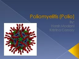
Slide2Polio (also called Poliomyelitis) is a highly infectious disease caused by a virus
The virus invades the nervous system and can cause permanent paralysis Polio is spread through person-to-person contact and can spread rapidly through a community
Most infected people (90%) have no symptoms or very mild symptomsHowever, one in 200 infections leads to permanent paralysis (can’t move parts of the body) and even death
What
is polio disease?
www.immune.org.nz
2
Slide3Poliomyelitis
The words polio (grey) and myelon (marrow, indicating the spinal cord) are derived from the Greek. It is the effect of poliomyelitis virus on the spinal cord that leads to the classic manifestation of paralysis.
3
Slide4The disease of poliomyelitis has a long history. The first example may even have been more than 3000 years ago. An Egyptian stele dating from the 18th Egyptian dynasty (1580 - 1350 BCE) shows a priest with a deformity of his leg characteristic of the flaccid paralysis typical of poliomyelitis.
.
First described by Michael Underwood in 1789
4
Slide5Introduction
A viral infection most often recognized by acute onset of flaccid paralysis.
Infection with poliovirus results in a spectrum of clinical manifestations from inapparent infection to non-specific febrile illness, aseptic meningitis, paralytic disease, and death.
Poliomyelitis is a highly infectious disease caused by three serotypes of poliovirus.
5
Slide6Two phases of acute poliomyelitis can be distinguished: a non-specific febrile illness (minor illness) followed, in a small proportion of patients, by aseptic meningitis and/or paralytic disease (major illness).
The ratio of cases of inapparent infection to paralytic disease ranges from 100:1 to 1000:1.
6
Slide7Outcomes of poliovirus infection
7
Slide8Epidemiological pattern
The epidemiological pattern of polio depends upon the degree of the socioeconomic development and health care services of a country. The pattern of the disease has been considerably modified by widespread immunization.
8
Slide9Causative organism
Poliovirus: belongs to “Picorna” viruses which are small RNA-containing viruses.
Polioviruses have three antigenically distinct types, giving no cross immunity:Type I: “Leon”; the commonest in epidemics
Type II: “Berlinhide”; the prevailing type in endemic areas.
Type III: “Lansing”; occasionally causes epidemics.
Polioviruses are relatively resistant and survive for a long time under suitable environmental conditions, but are readily destroyed by heat (e.g. pasteurization of milk, and chlorination of water).
9
Slide10Types of polioviruses
99% reduction
in cases of wild poliovirus since
1988
Type 1 (
341
cases
as of 20 November 2013†
)
Type 2 (eliminated worldwide in 1999)
Type 3 (none detected since November 2012)
Wild
Vaccine derived polioviruses (VDPV)
Most are circulating VDPVs (
cVDPVs
)*
~49-184 per year since 2008 (through 20 Nov 2013)
Type
2
cVDPVs
account
for
97%
of
cVDPVs
VDPVs*
Vaccine-associated paralytic poliomyelitis (VAPP)**
Estimated ~250-500 globally per year
Type
2
accounts
for about 40% of VAPP
VAPP**
†
More up-to-date numbers can be found at
http
://www.polioeradication.org/Dataandmonitoring/Poliothisweek.aspx
*Other extremely rare VDPVs include primary immunodeficiency VDPVs (
iVDPVs
) and ambiguous VDPVs (
aVDPVs
)
**Refers
to spontaneous reversion to
neurovirulence of one of the attenuated viruses in OPV. VAPP occurs in OPV recipients or their close contacts in contrast to cVDPVs which are widely transmitted in a community and are not likely to be related to contact with a recent vaccine recipient.
OPV related
10
Slide11Reservoir of infection
Man is the only reservoir of infection of poliomyelitis.
Man: cases and carriers
Cases: all clinical forms of disease
Carriers: all types of carriers (e.g. incubatory, convalescent, contact and healthy) except chronic type. In endemic areas, healthy carriers are the most frequent type encountered.
11
Slide12Foci of infection
Pharynx: the virus is found in the oropharyngeal secretions.Small intestine: the virus finds exit in stools.
12
Slide13Modes of transmission
Since foci of infection are the throat and small intestines, poliomyelitis spreads by two routes:
Oral-oral infection: direct droplet infection
Faeco
-oral infection:
Food-borne (ingestion) infection through the ingestion of contaminated foods. Vehicles include milk, water, or any others that may be contaminated by handling, flies, dust….Hand to mouth infection.
(polio virus has the ability to survive in cold environments. Overcrowding and poor sanitation provide opportunities for exposure to infection.)13
Slide14Poliovirus infection is
highly contagiousPoliovirus is spread mostly by the fecal-oral routePrimary mode of transmission – passage of the virus in stool to the mouth of another childCan also be spread through saliva or droplets from a sneeze or cough
How does poliovirus spread?
Virus transferred to objects from hands
Virus transferred to another child’s hands
Virus transferred ingested
Next cycle of infection
Child excretes virus in stool
14
Slide15Period of infectivity
Contact and healthy carriers: about 2 weeks
Cases: the cases are most infectious 7 to 10 days before and after the onset of symptoms. In the feaces, the virus is excreted commonly for 2 to 3 weeks, sometimes as long as 3 to 4 months.
In polio cases, infectivity in the pharyngeal foci is around one week, and in the intestinal foci 6-8 weeks.
Incubation Period: 7-14 days
15
Slide16Susceptibility
Age: more than 95% reported in infancy and childhood with over 50% of them in infancy.
Sex: no sex ratio differences, but in some countries, males are infected more frequently than females in a ratio 3:1.
Risk factors: (provocative factors of paralytic polio in individuals infected with polio virus): fatigue, trauma, intramuscular injections, operative procedures, pregnancy, excessive muscular exercise…
Immunity: The maternal antibodies gradually disappear during the first 6 months of life. Immunity following infection is fairly solid, although infection with other types of polio virus can still occur.
16
Slide17Sequelae of polio infection
Polio infection
Inapparent infection
Clinical poliomyelitis
Abortive polio
(minor illness)
Involvement of CNS
(major illness)
Paralytic polio
Non-paralytic
polio
Spinal polio
Bulbar polio
Bulbospinal polio
17
Slide18Inapparent infection
Incidence is more than 100 to 1000 times the clinical cases. No clinical manifestations, but infection is associated with acquired immunity, and carrier state.
18
Slide19Clinical poliomyelitis
Abortive polio (minor illness):
The majority of clinical cases are abortive, with mild systemic manifestations for one or two days only, then clears up giving immunity. Some abortive cases may be so mild to pass unnoticed.
Manifestations:
Moderate fever
Upper respiratory manifestations: pharyngitis and sore throatGastrointestinal manifestations: vomiting, abdominal pain, and diarrhea.
19
Slide20Clinical poliomyelitis (cont.)
II. Involvement of the CNS (major illness):
Affects a small proportion of the clinical cases, and appears few days after subsidence of the abortive stage. It takes two forms: nonparalytic and paralytic polio.
Nonparalytic polio is manifested by fever, headache, nausea, vomiting, and abdominal pain. Signs of meningeal irritation (meningism), and aseptic meningitis (pain and stiffness in the neck back and limbs) may also occur.
The case either recovers or passes to the paralytic stage, and here the nonpralytic form is considered as a “preparalytic stage”.
20
Slide21Clinical poliomyelitis (cont.)
Paralytic poliomyelitis:
Paralysis usually appears within 4 days after the preparalytic stage (around 7-10 days from onset of disease).
The case shows fever, headache, irritability, and different paralytic manifestations according to the part of the CNS involved, with destruction of the motor nerve cells, but not the sensory nerve cells.
Forms: spinal, bulbar, and bulbospinal.
21
Slide22Spinal polio
Different spinal nerves are involved, due to injury of the anterior horn cells of the spinal cord, causing tenderness, weakness, and flaccid paralysis of the corresponding striated muscles.
The lower limbs are the most commonly affected.
22
Slide23Bulbar polio
Nuclei of the cranial nerves are involved, causing weakness of the supplied muscles, and maybe encephalitis.
Bulbar manifestations include dysphagia, nasal voice, fluid regurgitation from the nose, difficult chewing, facial weakness and diplopia
Paralysis of the muscles of respiration is the most serious life-threatening manifestation.
23
Slide24Bulbospinal polio
Combination of both spinal and bulbar forms
24
Slide25Complications and case fatality
Respiratory complications: pneumonia, pulmonary edema
Cardiovascular complications: myocarditis, cor pulmonale.
Late complications: soft tissue and bone deformities, osteoporosis, and chronic distension of the colon.
Case fatality: varies from 1% to 10% according to the form of disease (higher in bulbar), complications and age ( fatality increases with age).
25
Slide26Case definition
The following case definition for paralytic poliomyelitis has been approved by CDC (1997)
Clinical case definition Acute onset of a flaccid paralysis of one or more limbs with decreased or absent tendon reflexes in the affected limbs, without other apparent cause, and without sensory or cognitive loss.
26
Slide27Case classification
Probable: A case that meets the clinical case definition.
Confirmed: A case that meets the clinical case definition and in which the patient has a neurologic deficit 60 days after onset of initial symptoms, has died, or has unknown follow-up status.
Confirmed cases are then further classified based on epidemiologic and laboratory criteria. Only confirmed cases are included in the
Morbidity and Mortality Weekly Report (MMWR)
.Indigenous case: Any case which cannot be proved to be imported.
Imported case: A case which has its source outside the country. A person with poliomyelitis who has entered the country and had onset of illness within 30 days before or after entry
27
Slide28HOT CASE
A case of AFP with any of the following set of conditions
-Age < 5 year plus H/O fever at onset plus asymmetrical proximal paralysis.
Age < 5 year with rapidly progressive paralysis leading to bulbar involvement (cranial nerves affected) & death.
Any case which in the opinion of SMO/DIO looks like polio.
28
Slide29Diagnosis
The diagnosis of paralytic poliomyelitis is supported by: (i) clinical course, (ii) virological
testing, and (iii) residual neurologic deficit 60 days after onset of symptoms. Laboratory testing, such as the measurement of antibodies (especially pre- and post-onset of paralysis), and other studies, such as magnetic resonance imaging, electromyography, and/or nerve conduction tests, can help strengthen or exclude the diagnosis of poliomyelitis.
29
Slide30WHO uses a sensitive screening case definition for the identification of AFP cases and for investigation of any case of AFP in a person younger than 15 years or in a person of any age in whom poliomyelitis is suspected.
However, virological examination is essential for confirmation of the diagnosis of poliomyelitis; this involves detection of poliovirus from the stools of patients with AFP and further characterization of the isolated poliovirus to determine whether it is vaccine-associated, vaccine-derived or wild virus.
30
Slide31Molecular diagnostics such as polymerase chain reaction are used to differentiate WPV, VDPV, and Sabin-like poliovirus.
In addition all discordant poliovirus isolates are partially sequenced to determine their origin and relatedness to other isolates. According to the laboratory results and review by national polio expert committees, cases are further classified as confirmed, polio-compatible, or polio-negative.
The AFP surveillance is supplemented by environmental surveillance which involves testing sewage or other environmental samples for the presence of poliovirus
31
Slide32Diagnosis and laboratory testing (cont.)
Serologic testing A four-fold titer rise between the acute and convalescent specimens suggests poliovirus infection.
Cerebrospinal fluid (CSF) analysis
The cerebrospinal fluid usually contains an increased number of leukocytes—from 10 to 200 cells/mm3 (primarily lymphocytes) and a mildly elevated protein, from 40 to 50 mg/100 ml.
32
Slide33Prevention
General prevention:Health promotion through environmental sanitation.
Health education (modes of spread, protective value of vaccination).
33
Slide34Prevention
Seroprophylaxis by immunoglobulins: Not a practical way of giving protection because it must be given either or before or very shortly after exposure to infection.
(0.3 ml/kg of body weight).
34
Slide35an armoury of five different vaccines to stop polio transmission:
Oral polio vaccine (OPV)Monovalent oral polio vaccines (mOPV1 and mOPV3)Bivalent oral polio vaccine (
bOPV)Inactivated polio vaccine (IPV)
35
Slide36prevention
Active immunization:Salk vaccine (intramuscular polio trivalent killed vaccine).Sabin vaccine (oral polio trivalent live attenuated vaccine).
36
Slide37Inactivated Polio Vaccine
Contains 3 serotypes of vaccine virusGrown on monkey kidney (Vero) cells
Inactivated with formaldehydeContains 2-phenoxyethanol, neomycin, streptomycin, polymyxin B
37
Slide38Oral Polio Vaccine
Contains 3 serotypes of vaccine virusGrown on monkey kidney (Vero) cells
Contains neomycin and streptomycinShed in stool for up to 6 weeks following vaccination
38
Slide39Salk versus Sabin vaccine
IPV (Salk)
OPV (Sabin)
killed formolised virus
Given SC or IM
Induces circulating antibodies, but not local (intestinal immunity)
Prevents paralysis but does not prevent reinfection
Not useful in controlling epidemics
More difficult to manufacture and is relatively costly
Does not require stringent conditions during storage and transportation. Has a longer shelf life.
live attenuated virus
given orally
immunity is both humoral and intestinal. induces antibody quickly
Prevents paralysis and prevents reinfection
Can be effectively used in controlling epidemics.
Easy to manufacture and is cheaper
Requires to be stored and transported at subzero temperatures, and is damaged easily.
39
Slide40Polio Vaccination of Unvaccinated Adults
IPVUse standard IPV schedule if possible (0, 1-2 months, 6-12 months)
May separate doses by 4 weeks if accelerated schedule needed
40
Slide41Polio Vaccine Adverse Reactions
Rare local reactions (IPV)No serious reactions to IPV have been documentedParalytic poliomyelitis (OPV)
41
Slide42What is vaccine-derived polio?
Vaccine-derived polioviruses (VDPVs) are rare strains of poliovirus that have genetically mutated from the strain contained in the oral polio vaccine.
42
Slide43VDPV can Cause Sustained Person-to-Person Spread and Reintroduction of Poliovirus
VDPVs are classified into 3 categories:Circulating VDPVs (cVDPVs)
Emerge in areas with inadequate OPV coverageMay lead to sustained person-to-person spread Immunodeficient-associated VDPVs (
iVDPVs
)
Isolated from persons with primary immunodeficiencies who exhibit prolonged VDPV infection after OPV exposure Potential source of PV reintroduction in the future; they may excrete virus for up to 20 years Ambiguous VDPVs (aVDPVs) Clinical isolates from persons with no known immunodeficiency orEnvironmental isolates with unidentified source
WHO. WER, 2006 . McLennan, et al. Lancet, 2000
Slide44VAPP: Rare But Serious
Vaccine-Associated Paralytic Polio:Defined as those cases of AFP from whose stool samples, vaccine-related poliovirus but no wild polio virus is isolated
Caused by mutation of vaccine virus during replication in the gut of vaccinee (reversion to neurovirulence)
VAPP is indistinguishable from naturally occurring polio
Same incubation period, range of severity and Case Fatality Rate
May affect both vaccinees & close contacts.
Sutter et al. Vaccines. 2008. Paul. Vaccine
, 2004 .John. Bull of the WHO
, 2004.44
Slide45B. Control of patient, contacts and the immediate environment:
Report to local health authority: Obligatory case report of paralytic cases as a Disease under surveillance by WHO, Class 1.
2) Isolation: Enteric precautions in the hospital for wild virus disease; of little value under home conditions because many household contacts are infected before poliomyelitis has been diagnosed.
3) Concurrent disinfection: Throat discharges, feces and articles soiled therewith. Terminal cleaning.
4) Quarantine: Of no community value.
45
Slide465) Protection of contacts: Immunization of familial and other
close contacts is recommended but may not contribute to
immediate control; the virus has often infected susceptibleclose contacts by the time the initial case is recognized.
6) Investigation of contacts and source of infection:
Occurrence
of a single case of poliomyelitis due to wild poliovirus must
be recognized as a public health emergency promptingimmediate investigation and planning for a large-scale response.
A thorough search for additional cases of AFP in thearea around the case assures early detection, facilitates
control and permits appropriate treatment of unrecognized
and unreported cases.
7) Specific treatment: None; however, Physical therapy is used to attain maximum function after paralytic poliomyelitis.
46
Slide47C. Epidemic measures:
In any country, a single case of poliomyelitis must now be considered a public health emergency, requiring an extensive supplementary immunization response over a large geographic area.
47
Slide48D. Disaster implications:
Overcrowding of non-immune groups and collapse of the sanitary infrastructure pose an epidemic threat.
48
Slide49E. International measures:
Poliomyelitis is a Disease under surveillance by WHO and is targeted for eradication by 2005.
National health administrations are expected to inform WHO immediately of individual cases and to supplement these reports as soon as possible with details of the nature and extent of virus transmission.
Planning a large-scale immunization response must begin immediately and, if epidemiologically appropriate, in coordination with bordering countries.
49
Slide50E. International measures (cont.):
Once a wild poliovirus is isolated, molecular epidemiology can often help trace the source.
Countries should submit monthly reports on case of poliomyelitis AFP cases and AFP surveillance performance to their respective WHO offices.
International travelers visiting areas of high prevalence must be adequately immunized.
50
Slide5151
Slide52Polio Eradication
Last case in United States in 1979Western Hemisphere certified polio free in 1994
Last isolate of type 2 poliovirus in India in October 1999Global eradication goal
52
Slide53WHY POLIO IS A CANDIDATE FOR ERADICATION ?
MAN IS THE ONLY RESERVIORNO LONG TERM CARRIER STATEROUTE OF TRANSMISSION IS FAECO-ORAL
HALF LIFE OF EXCRETED VIRUS IN SEWAGE SAMPLE IN TROPICAL CLIMATE LIKE INDIA IS 48 HOURS.POTENT AND EFFECTIVE VACCINE.
53
Slide54FOUR KEY STRATEGIES FOR POLIO ERADICATION
RI-PROGRAMME [ UIP ] MASS IMMUNIZATION(PPI) CAMPAIGNS
APF SURVEILLANCE MOPING UP IN FOCAL AREAS
54
Slide5555
Slide5656
Slide57Surveillance
Acute Flaccid Paralysis (AFP) surveillanceNationwide AFP (acute flaccid paralysis) surveillance is the gold standard for detecting cases of poliomyelitis.
57
Slide58The four steps of surveillance are:
finding and reporting children with acute flaccid paralysis (AFP)transporting stool samples for analysisisolating and identifying poliovirus in the laboratorymapping the virus to determine the origin of the virus strain.
58
Slide59WHY OPV ?
ALSO KNOWN AS SABIN VACCINEPOTENT LIVE VACCINEGIVES GUT IMMUNITYGIVES HERD IMMUNITY- INTERRUPT’s TRANSMISSION CYCLEEASY TO ADMINISTER
COST EFFECTIVE59
Slide60WHAT IS PULSE POLIO ?
TO IMMUNIZE ALL THE KIDS< 5YRS NATION WIDE ON A SINGLE DAY IN THE SHORTEST POSSIBLE TIME WITH OPV & THAT THE ENVIRONMENT WILL GET SATURATED WITH THE VACCINE VIRUS SO THAT IT WILL REPLACE THE WILD VIRUS AND THUS INTERUPT THE TRANSMISSION OF WILD VIRUS .
60
Slide61WHY AFP SURVEILLANCE INSTEAD OF
POLIO SURVEILLANCE ?
SURVEILLANCE OF A POLIO CASE ALONE IS NOT SUFFICIENT BECAUSE IT IS IMPOSSIBLEE TO PRECISELY IDENTIFY ALL CASES OF POLIO CLINICALLY DUE TO CONFUSING AND AMBIGUOUS CLINICAL SIGNS AND VARIABLE CLINICAL KNOWLEDGE & SKILLS OF DOCTOR.
CLINICALLY POLIO IN ACUTE STAGE, IS DIFFICULT TO DISTINGUISH FROM OTHER CAUSES OF ACUTE ONSET OF FLACCID PARALYSIS.-----
61
Slide62The principle of AFP surveillance is to identify children below 15 years with the syndrome of Acute Flaccid Paralysis
Acute-Rapid progression or short, brief duration Flaccid-Floppy or soft and yielding to passive stretching at any time during illness Paralysis–Loss of motor strength
Severe loss of motor strength is called paralysis or plegia
Paresis-indicates slight loss of motor strength
62
Slide63All cases of acute flaccid paralysis should be reported, irrespective of diagnosis, within 6 months of onset
All cases with flaccid paralysis should be reported and their stools must be collected within 14 days of onset. If it is not possible to collect stool specimens within 14 days, the specimens should still be collected up to 60 days after onset of paralysis.
63
Slide64Include every case with
: Current flaccid paralysis History of flaccid paralysis in the current illness Borderline or ambiguous clinical signs.
64
Slide65A case should not be included as AFP if
there is no evidence of acute flaccid paralysis at the time of examination or anytime during the course of illness or if the onset of paralysis is more than 6 months prior to notification.
65
Slide66Causes of AFP
Poliomyelitis
Gullain Barre Syndrome
Traumatic neuritis
Transverse
MyelitisAny other flaccid/lower motor presentation66
Slide67AFP SURVEILLANCE
STEPS
FOR EACH AFP CASECASE INVESTIGATION
2 STOOL SPECIMENS,COLLECTED 24 HOURS APART,AND WITHIN 14 DAYS OF ONSET OF PARALYSIS
SENT FOR CULTURES TO LAB TO ISOLATE POLIO VIRUS
ORI ACTIVITY & SEARCH FOR MORE AFP CASES IN THE AREA60 DAYS FOLLOW-UP EXAMINATION AFTER ONSET.
67
Slide68Onset of paralysis
Investigation of suspected case
(
≤
48 hours of report)
2 stool specimens collected (
≤
14 days since onset of paralysis)
24 hours apart
Outbreak response immunization additional case finding
60-day follow-up exam
Specimens arrive at national laboratory
Results reported from national laboratory
Poliovirus isolates send to regional reference laboratory for intratypic differentiation
Final classification of the case by the expert committee (
≤
12 weeks since onset of paralysis)
Flow diagram of case investigation,
stool specimen collection and
outbreak response immunization
≤ 3 Days
≤ 24 Days
≤ 7 Days
68
Slide69VIROLOGIC CLASSIFICATION SCHEME
NO WILD POLIOVIRUS
AFP
WILD POLIOVIRUS
INADEQUATE
STOOL SPECIMENS
TWO ADEQUATE*
STOOL SPECIMENS
NO RESIDUAL WEAKNESS
CONFIRM
COMPATIBLE
DISCARD
DISCARD
RESIDUAL WEAKNESS, DIED OR LOST TO F/U
DISCARD
EXPERT REVIEW
69
Slide70Surveillance indicators
Indicator
Minimum levels for certification standard surveillance
Completeness of reporting
At least 80% of expected routine reports should be received on time (inc. zero reports)
The distribution of reporting sites should be representative of the geography and demography of the country
Sensitivity of surveillance
At least one case of non-polio AFP should be detected annually per 100 000 population aged less than 15 years.
In endemic regions this rate should be two per 100 000.
Completeness of case investigation
All cases should have a full clinical and virological investigation with at least 80% of AFP cases having ‘adequate’ stool specimens collected.
Completeness of follow-up
At least 80% of AFP cases should have a follow-up examination for residual paralysis at 60 days after the onset of paralysis
Laboratory performance
All AFP case specimens must be processed in a WHO-accredited laboratory within the Global Polio Laboratory Network (GPLN)
70
Slide71Environmental surveillance
Environmental surveillance involves testing sewage or other environmental samples for the presence of poliovirus. Environmental surveillance often confirms wild poliovirus infections in the absence of cases of paralysis.
71
Slide72Systematic environmental sampling (e.g. in Egypt and Mumbai, India) provides important supplementary surveillance data.
Ad-hoc environmental surveillance elsewhere (especially in polio-free regions) provides insights into the international spread of poliovirus.
72
Slide73Certification of polio eradication
Certification is done for WHO Regions and not for individual countriesWHO Regions that have been certified polio free:Americas: 20 August 1994
Western Pacific: 29 October 2000Europe: 21 June 2002
73
Slide74Certification of a region is considered only when
All countries in the area demonstratethe absence of wild poliovirus transmission for at least three consecutive yearspresence of certification standard surveillanceglobal action plan for laboratory containment of wild poliovirus
74
Slide75Certification standard surveillance
Non-polio AFP rate: ≥2 per 100,000 population aged less than 15 years annuallyAdequate stool specimens : ≥80%All stool specimens tested for poliovirus at a WHO-accredited laboratory
Additional CriteriaInvestigation of AFP cases within 48 hours of initial notification: ≥80%
Timeliness of weekly AFP surveillance reports: ≥80%
75
Slide76Laboratory containment of WPV
To minimize the risk of reintroduction of WPV into the community from a laboratoryWHO action plan comprises three phases:Phase 1: laboratory survey and inventory
Phase 2: global certification: implement appropriate biosafety measuresPhase 3: post global certification: more stringent, will be prepared when there is global strategy to stop OPV
For regional certification evidence that phase 1 has been implemented
76
Slide77Rukhsar Khatoon last case of WPV detected in India (Jan 2011), her mother Shabida Bibi in Shahapar village, WB
77
Slide78Tommorows lecture
End game polio
78
Slide79Thank You
79