
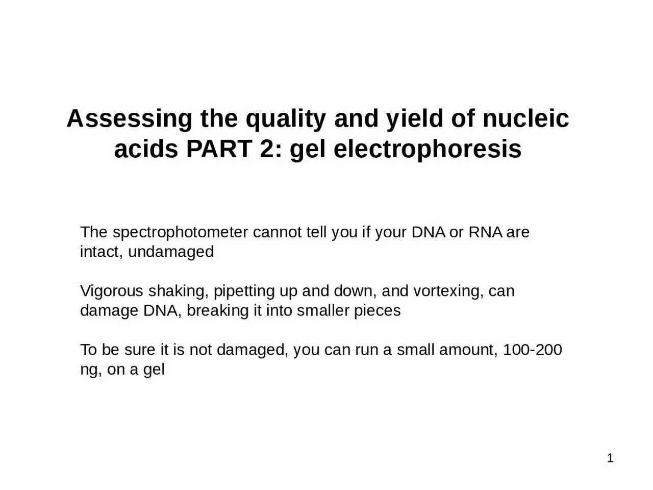
1 The spectrophotometer cannot tell you if your DNA or RNA are intact undamaged Vigorous shaking pipetting up and down and vortexing can damage DNA breaking it into smaller pieces To be sure it is not damaged you can run a small amount 100200 ng on a gel ID: 1046903
Download Presentation The PPT/PDF document "Assessing the quality and yield of nucle..." is the property of its rightful owner. Permission is granted to download and print the materials on this web site for personal, non-commercial use only, and to display it on your personal computer provided you do not modify the materials and that you retain all copyright notices contained in the materials. By downloading content from our website, you accept the terms of this agreement.


1. Assessing the quality and yield of nucleic acids PART 2: gel electrophoresis1The spectrophotometer cannot tell you if your DNA or RNA are intact, undamagedVigorous shaking, pipetting up and down, and vortexing, can damage DNA, breaking it into smaller piecesTo be sure it is not damaged, you can run a small amount, 100-200 ng, on a gel
2. Agarose gel electrophoresis for DNA:Gel electrophoresis separates DNA fragments (and RNA too) according to their size.DNA samples are loaded into wells (indentations) at one end of a gel, and an electric current is applied to pull them through the gel.DNA fragments are negatively charged, so they move towards the positive electrode (which is red in most systems). 2https://www.sciencedirect.com/topics/biochemistry-genetics-and-molecular-biology/agarose-gel-electrophoresis
3. Agarose gel electrophoresis for DNA:DNA fragments move different distances through a gel based on sizeWhen we speak of the “size” of a DNA fragment, we mean the length in base pairsAgarose is like a mesh with pores- smaller fragments can move through the pores more easily while longer fragments take longerA ladder is used to determine the sizes of the DNA fragments in each lane3https://www.sciencedirect.com/topics/biochemistry-genetics-and-molecular-biology/agarose-gel-electrophoresis
4. DNA runs through a gel in linear pieces proportionately to lengthRNA must be made linear to maintain the relationship between length and distance migrated When a gel is stained with a DNA-binding dye, the DNA fragments show up as bands, each representing a group of same-sized DNA fragments (same for RNA)4https://commons.wikimedia.org/w/index.php?title=File_talk:Gel_Electrophoresis.svg&action=edit&redlink=1
5. Dyes used to visualize DNA and RNA in gels (just a few common ones- there are others)Ethidium bromide: (UV)Fluoresces when bound to DNA with low background High sensitivityA known mutagenGel red: (UV) A safer form of ethidium, modified so that it cannot penetrate cell membranes, therefore should not be toxic (according to manufacturer)As sensitive as ethidium bromide (some claim it is more sensitive)More expensive than ethidium bromideSYBR safe: (blue light or UV)Safest of all these, because not toxic and UV is not neededLess sensitive than Gel redNot as pricey as Gel red5
6. Good quality DNA should migrate as a high molecular weight band, 20-40 kilobase pairs, kb), with little or no evidence of smearing.“ladder” of DNA fragmentswith known lengths providereferences to estimate the length of your DNA fragmentsIntact vs degraded DNAsmears of partially degradedDNAWe use a gel of 0.7- 1.5% for examining the length of DNA fragments and extent of DNA degradation.
7. Separation of RNA according to size by denaturing formaldehyde agarose gel electrophoresisMethod:Incubate RNA in the presence of formaldehyde and formamide at 55-65oC for a few minutes. (it is bad for you, use the fume hood)RNA denatures/ secondary structures are removedCarry out electrophoresis in agarose gel containing formaldehyde RNA remains denatured during electrophoresisRNases are denatured, inhibiting RNA degradationRNA molecules will migrate according to their molecular weight rather than ability to form secondary and tertiary structures*Nucleic acids must be LINEAR in order to migrate by size in a gel Caution:1. formaldehyde is a strong fixative/irritant2. buffer warms up during electrophoresis – fumes can fix your corneas!< <7
8. The integrity of ribosomal RNA can be used as an indicator of mRNA qualityRibosomal RNA (rRNA) makes up more than 80% of total RNA samples. Total RNA preps should display two prominent rRNA bands after gel electrophoresis. These correspond to the 25S and 18S rRNAs, in plants, 28S and 18s rRNAs in animals25S18S8Good quality RNA will have: - No/little evidence of smearing - 25S/18S or 28S/18S ratio ~ 2mRNA is present but in very small amounts, but will typically be intact if ribosomal RNAs are intact
9. SpeciesrRNASize (kb)Human18S1.928S5.0Mouse18S1.928S4.7Drosophila18S2.028S4.1Tobacco Leaf16S1.518S1.923S2.925S3.7Yeast18S2.026S3.8E. coli16S1.523S2.9Ribosomal RNA Sizes vary with organismDon’t memorize – you would need to know what sizes you are expecting to see for your research organismSvedberg (S) units9What are the 16S and 23S rRNAs in plant cells? Where do they come from?