
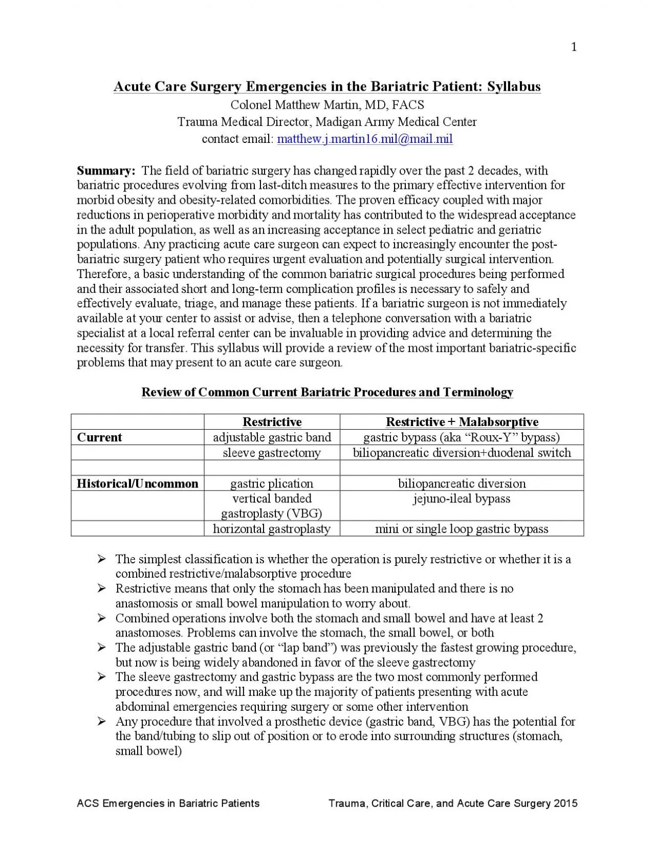
The sleeve gastrectomy and gastric bypass are the two most commonly performed procedures now and will make up the majority of patients presenting with acute abdominal emergencies requiring surgery or ID: 937295
Download Pdf The PPT/PDF document "Restrictive Malabsorptive Current adjus..." is the property of its rightful owner. Permission is granted to download and print the materials on this web site for personal, non-commercial use only, and to display it on your personal computer provided you do not modify the materials and that you retain all copyright notices contained in the materials. By downloading content from our website, you accept the terms of this agreement.


Restrictive + Malabsorptive Current adjustable gastric band gastric bypass (aka ÒRoux The sleeve gastrectomy and gastric bypass are the two most commonly performed procedures now, and will make up the majority of patients presenting with acute abdominal emergencies requiring surgery or some other intervention ! Any procedure that involved a prosthetic device (gastric band, VBG) has the potential for the band/tubing to slip out of position or to erod
e into surrounding structures (stomach, ¥ Leaks most commonly occur at the angle of His, just below or at the GE junction ¥ Entire anatomy still accessible by endoscopy abdominal abscess should be assumed to be due to a contained leak Key Points in the Evaluation of the Bariatric Emergency ! Most critical piece of history is identifying what prior procedure was performed; this will help guide the workup and areas of main concern on imaging st
udies (see Table 1 above) Patients manifesting with hemodynamic instability or signs of rapid progression of sepsis or clinical deterioration should be explored without undue delays or extensive workups ! Liberal use of imaging (fluoro, CT, or combined) to rule out major life ¥ Leaks may present as florid peritonitis and sepsis (uncontained) or with subacute symptoms of pain, fever, tachycardia, nausea/emesis (usually contained). ¥ There has been
a paradigm shift over the last 5 years away from routine operative exploration and repair for locally contained G-J leaks site hernia. " luminal J-J obstruction due to a formed hematoma can be a surgical emergency if completely obstructing " early obstruction of the J-J will cause both proximal dilation and emesis - these are both risk factors for disrupting the G-J anastomosis if not promptly treated by surgery or endoscopy " obstruction at or dis
tal to the J Although there are a number of signs on CT scan suggestive of an internal hernia, none are highly sensitive or specific. These include: " a mesenteric Òswirl signÓ indicating vascular torsion (arrow in Figure) is the most reliable sign " clustered loops of bowel in the left upper quadrant " small bowel loop behind the SMA " J-J anastomosis to the right of midline (should be on varies by anastomotic techniques and patient populations.
Common symptoms are epigastric pain with eating, but they can also present as spontaneous perforations. " the most common etiologies are smoking and NSAID use, but they can also be seen with large or dilated pouches that have more acid producing parietal cells " the majority should heal with acid suppression and smoking/NSAID cessation "ACS Emergencies in Bariatric Patients Trauma, Critical Care, and Acute Care Surgery 2015 II. Prior Sleeve Gastr
ectomy A. Early (within 30 days): Leaks and bleeding Endoscopic stenting to achieve adequate coverage of the leak is much more difficult compared to gastric bypass leaks, and should be referred to a very experienced endoscopic surgeon or gastroenterologist. ¥ Bleeding: Diagnosis can be made by a plain AP x-ray. The figure shows measurement of the Òphi angleÓ. This is the angle formed by a straight line through the long axis of the band and a v
ertical line "ACS Emergencies in Bariatric Patients Trauma, Critical Care, and Acute Care Surgery 2015 ¥ The 2 figures here show signs of a slipped band. ¥ Note the increased phi angle in the upper figure, now at almost 90 degrees relative to the vertical axis of the spine. ¥ An additional sign of slippage is when the appearance of the band on an AP xray changes from a solid disc (as seen in the upper figure) to a circular shape with the lumen vis
ualized (lower figure). ¥ This is known as the ÒO signÓ and should prompt immediate evaluation or intervention for slippage. ¥ The patient will usually present with acute intolerance to oral intake, persistent emesis and even difficulty with oral secretions. Although the diagnosis can be made based on the clinical picture and plain x-ray, a contrast swallow or CT scan can be helpful in equivocal cases or to delineate the degree of slippage and gast
ric herniation, and rule out a perforation. ¥ The ultimate treatment is removal of the band, but in many cases deflating the band by accessing the subcutaneous port and aspirating all fluid will relieve the symptoms and obviate the need for emergent surgery. This can also allow the band to slip back into normal position, but should still be followed by band removal or revision. Simplified Algorithm for the Bariatric Patient with Acute Abdominal C