
Director National Institute of Ayurveda Jaipur Raj 302002 Ph 9194180 79691 Email profsanjeevhpgmailcom sm This term has been used to describe a number of different anatomical abnormalities of the foot but over the years it has ID: 911073
Download Presentation The PPT/PDF document "CLUB FOOT (CTEV) Prof. Sanjeev Sharma" is the property of its rightful owner. Permission is granted to download and print the materials on this web site for personal, non-commercial use only, and to display it on your personal computer provided you do not modify the materials and that you retain all copyright notices contained in the materials. By downloading content from our website, you accept the terms of this agreement.


Slide1
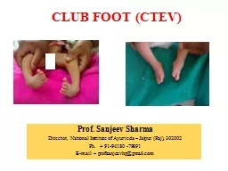
CLUB FOOT (CTEV)
Prof. Sanjeev Sharma
Director, National Institute of Ayurveda –
Jaipur (Raj), 302002Ph. + 91-94180 -79691E-mail – profsanjeevhp@gmail.com
sm
Slide2This term has
been used to describe a number of different anatomical abnormalities of the foot , but over the years it has
come to be synonymous
of the commonest congenital foot deformity i.e. CTEV. Club foot
Vague Term
Slide3Club Foot
Club foot is an Embryonic Malformation
Su.Su
. 24/6
Slide4Equinus
Derived from ‘ equine’ i.e. a horse who walk
s
on the toes . In this condition / deformity foot is fixed in a position of plantar flexion .
Slide5Calcaneus
Reverse of equinus.
Slide6Varus
The foot is inverted and adducted at the mid-tarsal joints so that sole faces inwards .
Slide7Valgus
The foot is everted and abducted at mid-tarsal joint.
Slide8Cavus
The logitudinal arch of foot is exaggerated.
Slide9Planus
The logitudinal arch of foot is flattened.
Slide10Splay
The transverse arch of foot is flattened .
Slide11Slide12Slide13Parts of the Foot
Hind Foot
Mid FootFore Foot
Slide14Bones of the Foot
Slide15CLUB FOOT
(CTEV)
Club foot is defined as a
structural deformity of the foot, characterised by fixed cavus and adductus of the forefoot, and varus and Equinus of the hind foot.
Slide16CLUB FOOT
(CONGENITAL TALIPES EQUINOVARUS)
It
is a congenital deformity 1/1000 births Involves one foot or both (>50% B/L).Male female ratio = 2:1First degree relatives = 2%Second degree relatives = 0.6%
In single foot involvement =
Rt
more than Lt.
Slide17Etiology
Idiopathic
Secondary
Slide18Idiopathic
Uterine Mechanical Factors
HereditoryOtogenic
/Arrest TheoryVascular HypothesisMusculo-ligamentous FibrosisPrimary Germ PlasmaArrest Fetal Development
Slide19In+Ad+Equate
Inversion at sub-talar joint
(In)
Adduction at talo-navicular joint and (Ad)Equinus at ankle joint, that is, a plantar flexed position, making the foot tend towards toe walking. (Equ
ate
)
Slide20Pathological Anatomy of the CTEV
Bones Involved:Talus
CuboidNavicularCalcaneum
Slide21The
Talus
and Calcaneus
are in severe flexion. The Calcaneus, Navicular and the Cuboid are adducted and inverted. The Navicular tuberosity is close to the medial malleolus.
The
Metatarsals
are
adducted.
calcaneus
Talus
Navicular
cuboid
Slide22CLUB FOOT – MORBID ANATOMY
First metatarsal in more plantar flexion than the lateral metatarsals
The entire foot is in supination
Forefoot is pronated in relation to the hind foot causing cavus
Slide23CLUB FOOT – MORBID ANATOMY
Firmly held in adduction and inversion by very tight ligaments and tendons in clubfeet
rotated medially in
relation to the talus
Calcaneus
Navicular
Cuboid
Slide24Muscles and TendonsLigaments
Joint Capsule
FasciaSkin
Vascular ChangesPathological Anatomy of the CTEV
Slide25Slide26Clubfoot
–Treatment Concepts
1. Conservative
French Method (Functional method) Kite’s Method (Serial plaster corrective casts)2. Surgical
Slide27Ponseti’s
Method
03.06.1914 – 18.10.2009
3. Conservative + minor surgery (If required)
Originally advocated by Ignacio
Ponseti
Slide284. Ayurvedic intervention +
Conservative +
minor surgery
Steps of this techniqueSnehana and Swedana before every manipulationEvery time manipulation followed by Above knee corrective cast.
Tenotomy
and cast after achieving full correction.
Finally daily
snehana
, manipulation and Brace application
Slide29Success of this technique depends on good plastering technique step by step
Ayurvedic
intervention + Conservative + minor surgery
Slide30Protocol of Corrective Procedure
1. Snehana / Swedana (10 min)
Su. Su. 25/3
Su. Sha. 10/15
Til
Tailam
or
Bala
Tailam
or
Tailam
suitable to tender skin of a newborn
Slide31Why Snehana and Swedana ??
Su. Chi. 4/7-8
Toning
of the skinIncrease in blood circulationRelaxation and toning of the muscles and ligaments
Slide322. Preliminary Manipulation
For 1 minute before POP to stretch soft tissues
Slide33The cavus is corrected by
extending
the first metatarsal and
supinating the forefoot.
Cavus
First metatarsal in more plantar flexion than the lateral metatarsals causing
Cavus
Slide34Front view. To initiate the correction of the clubfoot the first metatarsal is extended and the forefoot is held in supination in proper alignment with the midfoot and the calcaneus. In this position the foot can be abducted under the talus.
Cavus
Slide35Cavus
The forefoot must never be pronated
Slide36Varus and Adduction
To correct the deformity the foot distal to the talus must be made to rotate laterally, i.e
. abduct, underneath the talus which is fixed in the ankle mortise
Slide37The flexed foot, lightly supinated, is slowly abducted while counter pressure on the head of the talus stabilizes the bone against rotation in the ankle mortise. The medial tarsal ligaments are stretched allowing the calcaneus to abduct with the foot and the anterior tuberosity of the calcaneus is disengaged from its position under the head of the talus.
Slide38The lower part of the tibia is grasped by one hand with the index and middle
fingers resting on the inner aspect of the tibia just above the medial malleolus
where the markers are. The thumb rests on the lateral aspect of the head of the talus. The other hand grasps the forefoot and midfoot in slight supination. The clubfoot will be corrected when the dots on the head of the talus and on the navicular coincide and the anterior tuberosity of the calcaneus is engaged from its position under the head of the talus.
Slide39Complete correction of the clubfoot requires severe abduction of the midfoot and forefoot to stretch the tight medial tarsal ligaments.
Slide40Maintain correction by holding toes and applying counter pressure on talar head
Apply a thin layer of cotton padding
Cast Application
Slide41Removal of Plaster
(After 1 week) 5 days
Remove plaster just before new cast is to be given
Ask parents to clean the limb with soap and water
Slide42Again Snehana and Swedana
10 Min.
Manipulation 1-2 Min.
Re-application of cast in more corrected position
Slide43How Many Plasters?
Usually changed at weekly (5 days)
intervals6-8
(4-6) plaster applicationsUsually sufficient or till foot abducts to 700 abduction
Slide44The equinus is corrected by dorsiflexing the
fully abducted foot
A percutaneous tenotomy
of the Achilles tendon is often necessary to completely correct the equinus
Last plaster after
Tenotomy
for 3 weeks
Tenotomy
Slide45Serial photographs at weekly intervals of the correction
of
a severe clubfoot deformity in a 3-week-old infant.
A, At initial visit. B, After first cast. C, After second cast. D, After third cast. E, After fourth cast. F, Treatment result after percutaneous tendoachilles tenotomy.
Slide461
3
2
45
Slide47Denis Browne Brace
The shoes are worn for 23 hours a day for 3 months and are worn at night and during naps for up to 3 years.
Slide48Take Home Message
Most patients with clubfoot in early childhood can be treated by non-operative means
Ponseti’s
technique gives good and adequate correction and obviates the need for surgeryWhen Ayurvedic interventions like Mridu snehana and swedna is added it yields very good and early correction.
Ayurvedic doctors can practice this technique
Slide49T
hanks