
By Mira Younis 1 Definition Medically and scientifically death is not an event it is a process in which cellular metabolic processes in different tissues and organs cease to function at different rates ID: 1038691
Download Presentation The PPT/PDF document "Death & Post Mortem Changes" is the property of its rightful owner. Permission is granted to download and print the materials on this web site for personal, non-commercial use only, and to display it on your personal computer provided you do not modify the materials and that you retain all copyright notices contained in the materials. By downloading content from our website, you accept the terms of this agreement.


1. Death & Post Mortem ChangesBy: Mira Younis 1
2. DefinitionMedically and scientifically, death is not an event; it is a process in which cellular metabolic processes in different tissues and organs cease to function at different rates.The irreversible cessation of all integrated functioning of the human organism as a whole, mental or physical.legally defined as the irreversible cessation of function of 3 systems: (1) CNS (2) RS (3) CVSBefore the 1960’s, death was diagnosed only by cardio-pulmonary criteria: CNS criteria are new to the list.2
3. The 7 Processes of life 3DeathIs the absence of these 7 vital life processes.
4. DeathThis differential rate of cellular death has resulted in much debate – ethical, religious and moral – as to when ‘death’ actually occurs. The practical solution to this argument is to consider the death of a single cell (cellular death) and the cessation of the integrated functioning of an individual (somatic death) as two separate aspects.4
5. Types of death5
6. Somatic deathSomatic death is the death-- the permanent, irreversible death-- of an organism as a whole. It means that the individual will never again communicate or deliberately interact with the environment. The individual is irreversibly unconscious and unaware of both the world and their own existence. The key word in this definition is ‘irreversible’, as lack of communication and interaction with the environment may occur in a variety of settings such as deep sleep, under anaesthesia, under the influence of drugs or alcohol or as a result of a temporary coma.In human it is usually after brain death, as the other vital organs are unable to function without the brain. With modern technology, though, one can be brain dead but still have circulation and respiration artificially. In such a case one isn't somatically dead because other organs are still alive. Once artificial support is removed somatic death occurs, because the person is then entirely and completely inactive with regard to brain, circulation, and respiration.6
7. Cellular deathCessation of respiration (The utilization of oxygen) and the normal metabolic activity in the body tissues and cells. Cessation of respiration is soon followed by autolysis and decay, which, if it affects the whole body, is indisputable evidence of true death. The differences in cellular metabolism determine the rate with which cells die and this can be very variable – except, perhaps, in the synchronous death of all of the cells following a nearby nuclear explosion.Skin and bone will remain metabolically active and thus ‘alive’ for many hours and these cells can be successfully cultured days after somatic death.White blood cells are capable of movement for up to 12 hours after cardiac arrest. The cortical neuron, on the other hand, will die after only 3–7 minutes of complete oxygen deprivation. A body dies cell by cell and the complete process may take many hours.7
8. Clinical DeathThe 3 characteristics of clinical deathNo breathingNo circulationNo brain activity However, the most integral part which separates clinical death from somatic death, is that clinical death begins at the very onset of the symptoms of death, say right after cardiac arrest has cause the heart to stop. It lasts for about four minutes, and it is the interval in which life can be brought back through CPR. After a short few minutes, death is permanent.8
9. Brain DeathA brain deprived of oxygen survives for 3 to 7 minutes, making it the first organ to die when circulation or respiration ceases or is impeded, whatever the cause of trouble may be. After a few minutes, the brain can't be brought back to life by any means available today.This is brain death, and it's the reason why clinical death, the period in which a person can be resuscitated, is so short. Once the brain goes, the heart doesn't know how to pump and the lungs don't know how to breath.9
10. Indicators of cerebral deathUnconsciousness (coma)Absence of spontaneous breathing.Maximally dilated pupils which do not react to light.Absence of vestibulo-ocular reflexAbsence of corneal reflexAbsence of motor response to painful stimuliAbsence of gag reflex10
11. Apparent DeathA state of suspended animation that mimics death; it occurs in:ElectrocutionHypothermiaSun strokeDrowningDrug overdose (e.g. barbiturates)Head injurySuspended animation is the slowing of life processes by external means without termination!11
12. Diagnosing Death Is Important Medico-Legally :To detect the cause of death.To know the time of deathFor social reasonsFor organ donation For recognizing apparent deathFor statistical reasonsFor heritage reasons12
13. Mode vs Cause This is particularly important in relation to the documentary certification of deathsThe mode of death : an abnormal physiological state that pertained at the time of death: for example, 'coma', 'congestive cardiac failure', 'cardiac arrest' and 'pulmonary oedema'. These offer no information as to the underlying pathological condition and should not be used as the definitive cause of death unless further qualified by the more fundamental aetiological process.The cause: underlying pathological abnormality leading to that mode of death. For example:Hypertrophic cardiomyopathy (cause ) for Cardiac failure (mode)13
14. Certifying the Cause of DeathThe format for certifying the cause of death is now defined by the World Health Organization (WHO) and is an international standard that is used in most countries. The system divides the cause of death into two parts: the first part (Part I) describes the condition(s) that led directly to death; Part II is for other conditions, not related to those listed in Part I, that have also contributed to death. Part I is divided into 3 subsections (a), (b) and (c). These subsections are for disease processes that have led directly to death and that are causally related to one another, (a) being caused by or is a consequence of (b), which in turn is caused by or is a consequence of (c), etc. It is important to realize that, in this system of death certification, it is the disease lowest in the Part I list that is the most important, as it is the primary pathological condition in the ‘chain of events’ leading to death. Doctors should not record the mode of death (e.g. coma, heart failure) in isolation on the death certificate but, if a mode is specified, it should be qualified by indicating the underlying pathological abnormality leading to that mode of death. For example:Ia Cardiac failureIb Hypertrophic cardiomyopathyOrIa ComaIb Subarachnoid haemorrhageIc Ruptured congenital aneurysm.14
15. Manner of deathIn addition to the mode and cause of death, here is also the manner of death, which is not really a medical decision. Manner refers to the circumstantial events and is a legal categorization:Natural Accident Homicide Suicide Undetermined (2-5%) Pending Investigation 15
16. Post Mortem Changes16
17. Postmortem changes begin soon after death and progress along a timeline. Two processes, putrefaction and autolysis, begin to alter the body; either one may predominate, depending on the circumstances surrounding death, as well as the climate. Putrefaction involves the action of bacteria on the tissues of the body. This process, prevalent in moist climates, is associated with green discoloration of the body; gas production with associated bloating; skin slippage; and a foul odor.Autolysis is the breakdown of the body by endogenous substances. It proceeds most rapidly in organs such as the pancreas and stomach. It may predominate in more arid conditions and can eventually result in mummification.In most circumstances, autolysis and putrefaction occur in tandem. In temperate climatic conditions, they can result in rapid degradation of the tissues. These alterations may eventually produce great distortion of the body after death, hampering the interpretation of the postmortem findings but not ameliorating the value of the autopsy.17
18. Some of the more well-known postmortem changes, such as rigor mortis, livor mortis, and algor mortis, progress on a relatively set schedule; however, many external and intrinsic factors may affect their development. It should be remembered that the estimated period for the arrival and passage of these manifestations of the decomposition process is based on studies under very controlled conditions, including a temperate climate (ie, 75° F).In reality, many deaths occur outside of these “ideal” settings, and additional confounding variables may be present (eg, layered clothing, obesity, fever). Further, the longer the PMI, the less accurate the PMI estimate becomes.18
19. Body changes after deathInitially these changes can only be detected biochemically as the metabolism in the cells alters to autolytic pathways. Eventually the changes become visible and these visible changes are important for two reasons:Because a doctor needs to know the normal progress of decomposition so that he does not misinterpret these normal changes for signs of an unnatural death.Because they can be used in determining how long the individual has been dead (i.e the postmortem interval (PMI)).19
20. Post-mortem signs 20
21. Early Changes - EyeLoss of corneal and light reflexes.Mid-dilated pupils (1ry flaccidity) followed by constriction due to rigor mortis and finally dilaed again due to 2ry dilatation.Irregular size and shape of the pupils (anisocoria). The retinal vessels show the break up or fragmentation of the columns of blood, which is called ‘trucking’ or ‘shunting’ after 15 mins of death.Eyelids usually closed incompletelyTache noire : Where the sclera remains exposed to air, two black triangular spots appear at each side of the cornea (due to drying and accumulation of cellular debris).loss of intraocular tension within two hours, drops by half at time of death.21
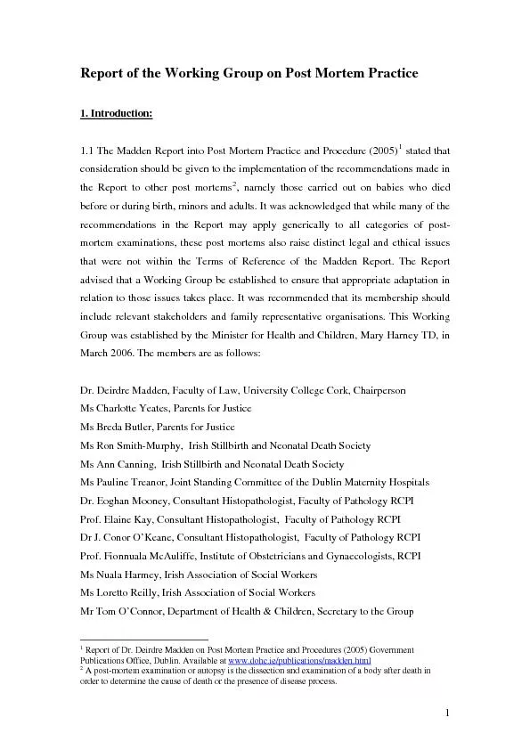
22. Tache Noire22
23. MusclesThe muscles rapidly become flaccid (primary flaccidity), with complete loss of tone, but they may retain their reactivity and may respond to touch and other forms of stimulation for some hours after cardiac arrest. Discharges of the dying motor neurons may stimulate small groups of muscle cells and lead to focal twitching, although these decrease with time.Loss of muscle tone in the sphincters may result in voiding of urine.23
24. SkinThe fall in blood pressure and cessation of circulation of the blood usually render the skin, conjunctivae and mucous membranes pale. Loss of elasticity (wounds do not gap)The skin of the face and the lips may remain red or blue in colour in hypoxic/congestive deaths. The hair follicles die at the same time as the rest of the skin and there is no truth in the belief that hair continues to grow after death, although the beard may appear more prominent against a pale skin.24
25. StomachRegurgitation is a very common feature of terminal collapse and it is a common complication of resuscitation.Gastric contents are identified in the mouth or airways in up to 25 % of all autopsies. The presence of this material cannot be used to indicate that inhalation was the cause of death unless it is supported by eyewitness accounts or by the microscopic identification of food debris in the peripheral airways.25
26. Body Cooling/ Algor MortisThe most useful indicator of time of death during the first 24 hours post-mortem.After death all metabolic activity ceases rapidly (muscles, liver) & circulation stops Heat production ceases soon after death.The body surface begins cooling immediately after death, followed by delay in deep organs cooling, until a heat gradient is set up between the core of the body and the surface. Delay “Temperature plateau” which can be variable from minutes to 2-3 hours.26
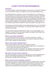
27. Rate of cooling27
28. In practice the temperature is either measured per rectum or intra-hepatic via an abdominal stab.The rate of body cooling:1°C/hr in summer.1.5°C/hr in winter.In winter it takes 18 hours to reach equilirium.Importance?Estimate time of deathEstimate the cause of death28
29. Hypostasis / Livor MortisIt’s the settling of blood in relatively lower parts of an organ or the body due to impaired or absent circulation Occurs after death, circulation of blood ceases & subsequent movement of blood is by gravity.Blood accumulates in the capillaries in the dependent parts of the body Purple or reddish purple discoloration of the adjacent skin.Within pressure areas such as the shoulder blades, buttock & calves Discoloration will be pale.Starts immediately after death.Apparent after 2 hrs. and fixed after 8 hrs.May not appear at all especially in infants, old and anemic or in those who have died from severe blood loss.29
30. Sites of hypostasisDepends on the position of the body before death:30SupineShoulders, buttocks.Heels pressing against surface giving a white color (pale).
31. Hanging Position.Distally in legs & feet.31Vertical
32. Chest, upper chest and upper limbs. 32DrowningDisposition of a body floating in water. Typically the head and limbs hang down, resulting in superficial injuries to the head/face, back of the arms and hands, knees and top of the feet.
33. As in epilepsy, drunken victims.Whitening around nose & lips. 33Face-downThe linear marks are formed by pressure from creases in the blanket. The pale areas around the mouth and nose are not necessarily signs of suffocation.
34. Other sites Heart: Mistaken for MI Lungs: Mistaken for pneumonia Intestine: Mistaken for hemorrhagic infarctionOnce hypostasis is established it has ability to undergo subsequent gravitational shift if the body is moved into a different posture. This is important because changes in the position of a body after the initial development of hypostasis will result in redistribution of the hypostasis and examination of the body may reveal two overlapping patterns.34
35. It can also be used by forensic investigators to determine whether or not a body has been moved (For instance, if the body is found lying face down but the pooling is present on the deceased's back, investigators can determine that the body was originally positioned face up).35
36. Color of HypostasisThe color of hypostasis is variable and depends on the state of oxygenation at death. It may be masked by dark skin colours, by jaundice or by some dermatological conditions.Colour changes that may act as indicators of possible causes of death:Cherry-pink: CO poisoning. Dark blue-pink: Cyanide poisoning.Brown: Methahemoglobinemia.Pallor: Anemia, hemorrhage (or normal in extremes of age).36
37. Timing and Permanence of HypostasisThe time is so variable that it has no significant role in determining the time of death.37Medico-legal Importance of HypostasisSure sign of death.Cause of death.Position before / after death.Indicate if the body was moved or not after death.
38. Hypostasis vs. bruises HypostasisBruises (Ecchymosis)Dependant areas Any whereWell defined edgesIll defined edgesBlood is retained in intact capillariesBlood escapes through ruptured capillariesSame level on surfaceRaisedPale over pressure areasRedIncision: blood flows from the cut vessel (washable)Incision: blood coagulates in tissue38With a bruise, blood will not flow from the cut
39. Rigor MortisTemperature-dependent, physico-chemical change that occurs within muscle cells as a result of lack of oxygen causing the limbs of the corpse to become stiff and difficult to move or manipulate.Death Cessation of respiration Depletion of oxygen Less ATP Secondary anoxic process Lactic acid cell cytoplasm becomes increasingly acidic With low ATP and high acidity, the actin and myosin fibres bind together and form a gel But Unlike normal muscle contractions, the body is unable to complete the cycle and release the coupling between the myosin and actin, creating a perpetual state of muscular contraction, until the breakdown of muscle tissue by digestive enzymes during decomposition.39
40. Rigor MortisR.M initiated when the ATP concentration falls to 85% of normal.It starts to develop about 2-3 hrs after death. Rigor develops uniformly throughout the body but it is first detected in smaller muscle groups such as those around the eyes, mouth, jaw & fingers.Peaks in the next 6-12 hrs.It concludes around 36-48 hrs after death.It resolves in the same order in which it develops.40
41. Timing of R.MEnvironmental temperature: Cold and wet slow onset, longer duration.Hot and dry fast onset, shorter durationMuscular activity before death: Muscles healthy and robust, at rest before death Slow onset, duration is longer.Muscles exhausted/ fatigued Onset is rapid, esp. in those limbs being used (E.g. in someone running at time of death, lower limbs develop RM faster than upper limbs).Increase activity (convulsions, electrocution, lightning) Rapid onset & short duration.Age: Extremes of age Rapid onset.41
42. Estimated time of death Body TMuscles’ toneETDWarm Flaccid<3 hrs.Warm Stiff 3-8 hrs.Cold Stiff 8-36 hrs.Cold Flaccid >36 hrs.42 A crude but useful aide-memoire.
43. Rigor Mortis (cont’d)R.M in Iris: May affect the eyes unequally, making the pupils unequal.R.M in the Heart: Contracted, stiff LV may be mistaken for LV hypertrophy.R.M in Dartos muscle of scrotum: Rigor in Dartos constricts testes and epididymis expulsion of semen.R.M in Erector Pilli muscles attached to hair follicles: Goose bumps, hair stands up.43
44. How to test for Rigor?It is best to test for rigor across a joint using very gentle pressure from one or two fingers only; the aim is to detect the presence and extent of the stiffness, not to ‘break’ it.If rigor is broken by applying too much force, those muscle groups cannot reliably be tested again.44
45. Cadaveric SpasmAlso known as instantaneous rigor or rigidity, or cataleptic rigidity.Rare form of muscular stiffening that occurs at the moment of death, persists into the period of rigor mortis and can be mistaken for rigor mortis.The cause is unknown, but usually associated with violent deaths happening with intense emotion.May affect all muscles in the body, but typically only groups, such as the forearms, or hands.Maybe seen in cases of drowning victims when grass, weeds, roots or other materials are clutched, and provides proof of life at the time of entry into the water.Often demonstrates the last activity one did prior to death and is therefore significant in forensic investigations, e.g. clinging on a knife tightly.45
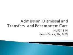
46. Cadaveric spasm in a drowning victim: This victim grasped at some ivy as he fell into water.Victim of suicide: The cadaveric spasm has maintained the position of his arms after the shotgun has been removed.Cadaveric Spasm46
47. Rigor Mortis Cadaveric SpasmOnset delayed after death (2-3 hrs.).Duration up to 36 hrs.Onset is instantaneous.Duration is a few hours, until it is replaced by rigor mortis.Intensity comparatively moderate.Intensity comparatively very strong.Mechanism of formation: Breakdown of ATP below critical level.Mechanism of formation unknown, but predisposing factors: Excitement, fear, fatigue, exhaustion, nervous tension, contraction of M’s at time of death.All muscles of the body are affected gradually.Selected muscles, which were in a state of contraction at the time of death, are affected.47
48. Conditions Mistaken as R.MHeat stiffness:Exposure of a body to intense heat (burning, high voltage electrocution, etc.) Coagulation of muscular proteins Muscular shortening.Cold stiffness:Exposure of the body to extreme cold (<-5⁰C) Solidification of subcutaneous fat and muscles, freezing of synovial fluid in joints.Rigor mortis halted until thawing occurs, after which it develops very rapidly.48
49. Medico-legal Importance of R.MMay help in time estimation.May help in finding the cause of death.May help to know the position.Sure sign of death.49
50. Post-Mortem DecompositionIn the cycle of life, dead bodies are usually returned, through reduction into their various components, to the chemical pool that is the earth. Some components will do this by entering the food chain at almost any level – from ant to tiger – whereas others will be reduced to simple chemicals by the autolytic enzymic processes built into the lysosomes of each cell.Putrefaction.Mummification.Adipocere.Skeletelization.50
51. PutrefactionThe normal final sign of death.Starts immediately after death at the cellular level. Becomes visible in 48-72 hrs.Two phenomena for putrefaction:Autolysis: Occurs by digestive enzymes released from the cells after death.Bacterial action: Most of them come from the bowel and Clostridium predominates (same bacteria that causes gas gangrene).The 1st visible sign of putrefaction is green or greenish red discoloration of the skin of the anterior abdominal wall. Normally starts in the right iliac fossa.51
52. PutrefactionThe blood vessels provide an excellent channel for bacterial spread throughout the body Decomposition of Hb which, when present in the superficial vessels, results in linear branching patterns of brown discoloration of the skin that is called ‘marbling’.As the superficial layers of the skin lose cohesions, blisters full of red or brown fluid form in many areas. When the blisters burst, the skin sloughs off. Considerable gas formation is common and the body begins to swell, with bloating of the face, abdomen, breasts and genitals.52
53. Marbling 53
54. PutrefactionThe increased internal pressure causes the eyes and tongue to protrude and forces bloody fluid up from the lungs and it will often leak out of the mouth and nose as ‘purge’.In general terms, within a week or so the body cavities will burst and the tissues will liquefy and drain away into the underlying ground.Brain & epithelial tissues are the 1st to be affected by putrefaction. Heart, uterus & prostate may survive for longer periods.54
55. Influences on PutrefactionA high environmental humidity will enhance putrefaction. Bodily habits of the decedent; obese individuals putrefy more rapidly than those who are lean. Putrefaction will be delayed in deaths from exsanguination (bleeding to death) because blood provides a channel for the spread of putrefactive organisms within the body. Conversely, putrefaction is more rapid in persons dying with widespread infection, congestive cardiac failure or retention of sodium and salts. 55
56. Influences on PutrefactionAge: more rapid in children than in adults, but the onset is relatively slow in unfed new-born infants because of the lack of commensal bacteria. Heavy clothing and other coverings, by retaining body heat, will speed up putrefaction. Rapid putrefactive changes may been seen in corpses left in a room which is well heated.Injuries to the body surface promote putrefaction by providing portals of entry for bacteria and the associated blood provides an excellent medium for bacterial growth. 56
57. MummificationA body lying in dry and warm conditions, either climatic or in the microenvironment, may desiccate instead of putrefying.Drying & shriveling of the tissues.Brown in color.The natural mummification process usually happens in extremely dry environments that allow the fast dehydration of tissues, simultaneously slowing down or inhibiting the decomposition by bacteria and other microorganismsAlso seen in newborn infants (sterile) whose bodies are placed in cool dry environments.No growth of micro-organisms.57
58. 58The time required for complete mummification can’t be precisely stated but it takes several weeks to months, depending on the size of the body (More likely in the thin individual) and atmospheric conditions.Once the changes are complete, the body will remain in that condition indefinitely.Mummification is partial, as 25% of body weight is preserved.Other factors affecting mummification:age (it is more common in newborns) gender (occurs more often in female) cause of death (large hemorrhages,ante-mortem prolonged administration of antibioticspoisoning by arsenic and potassium cyanideMummification
59. 59Medicolegal Importance of MummificationCause of Death.Can detect abnormal pathology inside deep organs.
60. AdipocereAlso known as "grave wax," adipocere (from the Latin, adipo for fat and cera for wax)is a grayish-white postmortem matter caused by fat decomposition, which results from hydrolysis and hydrogenation of the lipids (fatty cells) that compose subcutaneous fat tissues. Moisture is necessary.The optimum conditions for the formation of adipocere:Wet, warm environment (Sometimes original body water being sufficient for adipocere).Bacterial activity (C. perfiringes).It occurs in:Subcutaneous fat of the cheeks ,breast, buttocks.May occur in internal organs such as liver, kidney & heart. It needs months to occur, and occurs partially.60
61. 3 stagesIn early stages: Adipocere is a pale, rancid, greasy semi-fluid material with a most unpleasant smell. Later: Becomes more brittle and whiter.When fully formed, adipocere is a grey, firm, waxy compound which maintains the shape of the body.61Adipocere formation in an infant buried for 3 years. Thebody fat has been converted into brittle waxy material, which formsa shell around the skeleton.
62. 62
63. Medico-legal Importance of AdipocerePreserve the body which can permit identification after death.It may give conclusions about the cause of death.It indicates that the time interval since death was at least weeks to several months.63
64. Immersion and burialImmersion in water or burial will slow the process of decomposition.Body in air will decompose twice as fast as a body in water and four times as fast as a body under the ground.The first change that affects the body in water is the loss of epidermis. Gaseous decomposition progresses and the bloated body is often lifted to the surface by these gases, most commonly at about 1 week but this time is extremely variable. 64
65. SkeletelizationThe environment is more important than the time in this process.12-18 months: Soft tissues will be absent.Tendons, ligaments, hair and nails will be identifiable for some time after that.After 3 yrs: the bones will be bare and disarticulated.In temperate zones the bones will remain solid & heavy with the preservation of bone marrow in long bones for a number of years, that can sometimes be suitable for specialist DNA analysis.After 40-50 years:Bone surface becomes dry & brittle.Marrow cavity will be empty.65
66. Estimating the Time of DeathUnfortunately, all methods now in use to determine the time of death are to a degree unreliable and inaccurate. They usually give vague or answers. The longer the postmortem interval, the less precise the estimate of the interval.66
67. Estimating the Time of DeathCore body temperature:The best and the most commonly used.Rigor mortis.Hypostasis: Complete after 6 hrs.Chemical changes in vitreous.all electrolytes in the body change after death except K. There is linear relation b/w K level and time passed after death up to 120 hrs. Measured from vitrous humorWhen there is high urea concentration, there is an electrolyte imbalance and K can no longer be used as an indicatorMeasurement of potassium levels in the eyes can vary greatly from left to right eyes in the same corpse in ideal conditions.From a medico-legal standpoint, this technique is frowned upon, and so is the most infrequently used method to determine post mortem interval.Eye pressure: Eye balls become softer, and less fluid pressure in the first 3 hrs.67
68. Estimating the Time of DeathGastric emptying: Depend on type of meal and emotional status.The entomology of dead:Studying insects & their maggots which infest the dead body for estimating the probable time of death.Different types of insects infest the dead body at different stages after death occurs.Scene markersThough unscientific, is often more accurate than determinations made by scientific means.68
69. 69THANK YOU