

and Function Bacterial Form and Function A Structures common to all bacterial cells 1 Cell membrane 2 Cytoplasm 3 Ribosomes 4 One or a few chromosomes Bacterial Form ID: 805965
Download The PPT/PDF document "Bacteria and Archaea Bacterial Form" is the property of its rightful owner. Permission is granted to download and print the materials on this web site for personal, non-commercial use only, and to display it on your personal computer provided you do not modify the materials and that you retain all copyright notices contained in the materials. By downloading content from our website, you accept the terms of this agreement.


Slide1
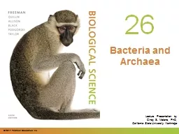
Bacteria
and
Archaea
Slide2Bacterial Form
and
Function
Bacterial
Form
and Function
A
. Structures common to
all
bacterial cells
1
. Cell membrane
2
. Cytoplasm
3
. Ribosomes
4
. One (or a few)
chromosomes
Slide3Bacterial Form
and Function
B. Structures found in
most
bacterial cells
1
. Cell wall
2
. Surface coating or
glycocalyx
C
. Structures found in
some
bacterial cells
1
. Flagella
2
.
Pili
Slide4Bacterial Form
and Function
3. Fimbriae
4
. Capsules
5
. Slime layers
6
. Inclusions
7
. Actin cytoskeleton
8
.
Endospores
Slide5Slide6Shapes & Arrangements
D. Bacterial Arrangements and
Sizes
1. Three
general shapes
A)
Coccus
– roughly spherical
B)
Bacillus – rod-shaped
Coccobacillus
– short and plump
Vibrio
– gently curved
C)
Spirillum
–
curviform
or spiral-
shaped
Slide7Shapes & Arrangements
2
.
Pleomorphism
– when cells of a single species vary to some extent
in
shape and size
3
. Arrangements & Groupings
A)
Cocci
– greatest variety in arrangement
1) Singles
2)
Pairs (
diplococci
)
Slide8Shapes & Arrangements
3
)
Tetrads
4
)
Irregular clusters (staphylococci)
5
)
Chains (streptococci)
6
)
Cubical packet (
sarcina
)
Slide9Shapes & Arrangements
B)
Bacilli – less varied
1) Singles
2)
Pairs (
diplobacilli
)
3
) Chains
(
streptobacilli
)
4
)
Row of cells oriented
side-by-side
(palisades
)
Slide10Shapes & Arrangements
C)
Spirilla
1) Usually singles
2) Occasionally
found in short
chains
Slide11External Structures
External
Structures
A
. Appendages: Cell extensions
1
. Common but not present on all species
2
. Can provide motility (
flagella,
pili
and axial filaments)
3
. Can be used for attachment and “mating” (
pili
and fimbriae
)
Slide12External Structures
4. Flagella
A) Three parts: Filament, hook (sheath), and basal body
1
) Filament
a
) Whip-like, helical structure
2
) Hook
a
) Holds the filament
b
) Attached to the rod portion of the basal
body
Slide13External Structures
3) Basal body
a
) A complex structure consisting of a rod, 4 rings and
a
motor contained within the cell envelope
b
) Activation of the motor causes the hook (and
therefore,
the filament) to
swivel
Slide14Figure 4.2
Slide15External Structures
B) Vary in both number and
arrangement
1
)
Monotrichous
– single flagellum
2
)
Lophotrichous
– small bunches or tufts of flagella
emerging
from the same site
3
)
Amphitrichous
– flagella attached at both ends of the cell
4
)
Peritrichous
– dispersed randomly over the structure
of the cell
Slide16Figure 4.3
Slide17External Structures
C)
Flagellar
Function
1
)
Chemotaxis
– movement of the cell in response to a
chemical
signal
a
) 2 types
i
) Positive
chemotaxis
ii
) Negative
chemotaxis
Slide18External Structures
b) Some photosynthetic bacteria exhibit
phototaxis
c
) Move is accomplished through a
series of
runs
and
tumbles
i
) Run – linear movement
(
a) Created by counterclockwise
flagellar
rotation
Slide19External Structures
ii) Tumble –
cell
stops and reverses directions or
spins
in place
(
a) Created by clockwise
flagellar
rotation
Slide20Slide21External Structures
5. Axial Filaments
A
) Also known as a
periplasmic
flagella
B
) Seen in a special group of bacteria known as
spirochetes
C
) Consists of a filament and hook but the entire structure is
located
between the cell wall and membrane (the
periplasmic
space)
D
) Creates movement through twisting and flexing
actions
Slide22External Structures
6.
Pili
A
)
Elongated,
rigid
hollow structures
B
) Found on some gram-negative bacteria
C
) Involved in attachment, movement and conjugation
7
. Fimbriae
A
) Small,
bristle-like
fibers
B
) Tend to stick to each other and to
surfaces
Slide23External Structures
8.
Glycocalyx
A) Develops
as a coating of
repeating polysaccharide
units,
protein
, or both
B)
Differ among bacteria in thickness, organization, and
chemical composition
Slide24External Structures
1) Slime layer – a loose shield that protects some bacteria
from
loss of water and nutrients
2
) Capsule – when the
glycocalyx
is bound more tightly to
the
cell and is denser and
thicker
Slide25Figure 4.9
Slide26External Structures
C
) Functions of the Glycocalyx
1
) Protects the cell
a
) Formed by many pathogenic bacteria to protect the
bacteria
against phagocytes
2
) Sometimes helps the cell adhere to the environment
a
) Important in formation of biofilms
3
) Helps prevent the loss of water and nutrients
Slide27The Cell Envelope
The
Cell Envelope:
The
Boundary layer of Bacteria
A. Majority
of bacteria have a cell envelope
B. Composed
of two or three basic layers
1
. Cell wall
2
. Cell membrane
3
. In some bacteria, the outer
membrane
Slide28The Cell Envelope
C
.
Differences in Cell Envelope Structure
1
. The differences between gram-positive and gram-negative
bacteria
lie in the cell envelope
2
. Gram-positive
A
) Two layers – cell wall and
cell membrane
3
. Gram-negative
A
) Three layers – outer membrane, cell wall, and
cell membrane
Slide29The Cell Envelope
D
.
Structure of the Cell Wall
1
. Helps determine the shape of a bacterium
2
. Provides strong structural support
3
. Most are rigid because of
peptidoglycan
content
4
. Keeps cells from rupturing because of changes in pressure
due to osmosis
Slide30The Cell Envelope
A) Target of many antibiotics – disrupt the cell wall, and
cells have
little protection from lysis
5
. Gram-positive cell wall
A
) A thick sheath of
peptidoglycan
B
) There is little space between the cell wall and membrane
(
periplasmic space
)
Slide31The Cell Envelope
C) 2 molecules (besides peptidoglycan) are commonly found
1
)
T
eichoic
acid – binds together layers of peptidoglycan
2
)
L
ipoteichoic
acid – link the peptidoglycan layers to the
cell
membrane
D
) Gram positive cells walls are less susceptible to lysis
(
stronger) but more permeable than gram negative
bacteria
Slide32The Cell Envelope
6. Gram
-negative
c
ell
w
all
A
) Single, thin sheet of peptidoglycan
B
) A wide periplasmic space surrounds the peptidoglycan
C
) Unlike gram positive bacteria, it possesses an outer
membrane
(a.k.a. LPS layer)
1
) Similar to the cell membrane, except it contains
specialized
polysaccharides and
proteins
Slide33The Cell Envelope
2) Innermost layer – phospholipid layer anchored by
lipoproteins
to the peptidoglycan layer below
3
) Outermost layer – contains lipopolysaccharide: 2
important
components
a
) Lipid A – found within the bilayer; recognized by our
immune systems
Slide34The Cell Envelope
b) O-specific polysaccharide – found externally; used to
identify
certain strains/species of bacteria (
E. coli
O157
:H7)
4
) Outer membrane serves as a partial chemical sieve
a
) Only relatively small molecules can
penetrate
Slide35Figure 4.14
Slide36The Cell Envelope
D) Gram negative bacteria are less permeable (because of the
LPS
) but more susceptible to lysis than gram positive
bacteria
E
.
Cell Membrane Structure
1
. Also known as the cytoplasmic membrane or plasma membrane
2
. Contain primarily phospholipids and
proteins
Slide37The Cell Envelope
3. Functions
A
) Provides a site for functions such as energy reactions,
nutrient
processing, and synthesis
B
) Regulates transport (selectively permeable membrane)
C
)
Secretion
Slide38Internal Structures
Bacterial
Internal Structure
A
. Contents of the Cell Cytoplasm
1
. Gelatinous solution
2
. Site for many biochemical and synthetic activities
3
. 70%-80% water
4
. Also contains larger, discrete cell masses (
chromosome/nucleoid, plasmids
, ribosomes, inclusions, and actin strands
)
Slide39Internal Structures
5.
Bacterial Chromosome
A
) Single circular strand of
essential DNA
B
) Aggregated in a dense area of the cell – the nucleoid
6
. Plasmids
A
) Extra, nonessential pieces of DNA
B
) May be found floating freely in the cytoplasm or attached to
the chromosome
Slide40Internal Structures
C) Often confer protective traits such as drug resistance or the
production
of toxins and enzymes
D
) Can be transferred from one bacterium to another naturally
or
artificially, thereby transferring the traits it
carries
Slide41Internal Structures
7
.
70S Ribosomes
A
) Made of
rRNA
and protein
B
)
The
site of protein production in the
cell
Slide42Internal Structures
8. Inclusions – also known as inclusion bodies
A
) Some bacteria lay down nutrients in these inclusions during
periods
of nutrient abundance
B
) Serve as a storehouse when nutrients become depleted
1
) Some enclose condensed, energy-rich organic substances
C
) Some aquatic bacterial inclusions include gas vesicles to
provide
buoyancy and
flotation
Slide43Internal Structures
9. Actin Cytoskeleton
A
) Long polymers of actin
B
) Contribute to cell
shape
Slide44Slide45Internal Structures
B. Bacterial Endospores: An Extremely Resistant Stage
1
. Dormant bodies produced by
Bacillus, Clostridium,
and
Sporosarcina
2
. These bacteria have a two-phase life cycle
A
) Phase One: Vegetative cell
1
) Metabolically active and
growing
Slide46Internal Structures
2) Can be induced by the environment to undergo spore
formation
(sporulation)
B
) Phase Two: Endospore
1
) Stimulus for sporulation – the depletion of nutrients
a
) Process takes 6-8 hours
2
) Vegetative cell undergoes a conversion to a sporangium
3
) The DNA of the cell is
duplicated
Slide47Internal Structures
4) A septum forms dividing the cell into unequal parts each
with
its own DNA
5
) The larger portion engulfs the smaller portion resulting
in
a
forespore
6
) A thick peptidoglycan coat forms around the
forespore
making
it impervious to other substances and heat
resistant
; it is now an
endospore
Slide48Internal Structures
7) The endospore is released as the sporangium
deteriorates
8
) The endospore remains dormant until conditions
improve
around
it
Slide49Slide50Internal Structures
C) Endospores are the hardiest of all life forms
1
) Withstand extremes in heat, drying, freezing, radiation,
and
chemicals
a
) Resist ordinary cleaning methods
2
) Some viable endospores have been found that were more
than
250 million years
old
Slide51Internal Structures
D) Germination
1
) Breaking of dormancy
2
) In the presence of water and a specific germination agent
the
spore will break down and a vegetative cell will
develop
3
) Quite rapid (1 ½ hours
)
Slide52Internal Structures
E) Medical Significance
1
) Most endospore-forming bacteria are relatively harmless
but
with some bacteria the endospore play a vital role in their
pathogenicity
a
) Examples include
Bacillus anthracis, Clostridium
tetani
,
Clostridium perfringens
, and
Clostridium
botulinum
Slide53Classification Of Bacteria
Classification
Systems
for Bacteria
and Archaea
A. Introduction – There are multiple criteria by which you can classify an organism including
shape
, variations in arrangement, growth characteristics, and habitat
1
. Metabolic Activities (Carbon, Energy & Oxygen sources)
A
) Recall that microbes may vary in their carbon & energy
sources
Slide54Classification Of Bacteria
1)
Phototrophs
– use light energy to extract
carbon
a) Photoautotrophs– obtain carbon from inorganic compounds (i.e. CO
2
)
b)
Photoheterotrophs
– obtain carbon from organic compounds (i.e. glucose
)
Slide55Classification Of Bacteria
2
)
Chemotrophs
– use chemical energy to extract carbon
a
)
Chemoheterotrophs
– obtain carbon from organic compounds (i.e. glucose)
b
)
Lithoautotrophs
– obtain carbon from inorganic compounds (i.e. CO
2
)
Slide56Classification Of Bacteria
B) Recall that microbes also vary in their oxygen requirements
1
) Aerobes – use oxygen as their final electron acceptor in metabolism
2
) Anaerobes – do not use oxygen as their final electron acceptor; often use sulfate,
nitrate
, carbonate or pyruvate; some cannot survive in the presence of
oxygen
Slide57Classification Of Bacteria
2.
Ecophysiology
(preferred environment)
A
) Microbes also vary by their preferred habitat
1
) Some microbes thrive in terrestrial environments
2
) Some microbes thrive in aquatic environments
3
) Some microbes thrive on or within animals
4
) Some microbes thrive in extreme
conditions
Slide58Classification Of Bacteria
3. Movement
A
) A small number of bacteria are unique in their mode of motility
B. Species and Subspecies
1
. Common definition of species used for animals (can produce viable offspring only
when
it mates with others of its own kind) does not work for bacteria
A
) Bacteria do not exhibit a typical mode of sexual
reproduction
Slide59Classification Of Bacteria
2. For bacteria – a species is a collection of bacterial cells, all of which share an overall
similar
pattern of traits
A
) Individual members of a bacterial species can show variations
B
) Subspecies, strain, or type – bacteria of the same species that have differing
characteristics
C
) Serotype – representatives of a species that stimulate a distinct pattern of antibody
responses
in their
hosts
Slide60Obligate Intracellular Parasites
Survey
of
Bacterial Groups
with Unusual Characteristics
A. Unusual Forms of Medically Significant Bacteria
1
. Obligate Intracellular Parasites
A
)
Rickettsias
(Gram negative; multiple shapes usually
cocci
)
1
) Most-pathogens that alternate between a mammalian host and blood-sucking
arthropods
Slide61Obligate Intracellular Parasites
2) Cannot survive or multiply outside a host cell
3
) Human diseases:
a
) Rocky Mountain Spotted Fever by
Rickettsia
rickettsii
b
) Endemic typhus by
Rickettsia
typhi
c
) Epidemic typhus by
Rickettsia
prowazekii
Slide62Obligate Intracellular Parasites
B)
Chlamydias
1
) Genera
Chlamydia
and
Chlamydophila
2
) Require host cells for growth and metabolism
3
) Human diseases
a
)
Chlamydia trachomatis
– causes a severe eye infection and the
STD
Slide63Spirochetes
2.
Bacteria that
Move by Unusual Mechanisms
1
) Spirochetes (Gram negative
spirillum
) move via an axial filament
a
) Axial filament – sets of flagella found at the poles of the bacteria and located
within
the
periplasm
b
) Cell moves in a corkscrew
fashion
Slide64Spirochetes
c) Examples include:
i
)
Treponema
sp., which causes syphilis
ii
)
Borellia
sp., which causes Lyme
disease
Slide65Photosynthetic Bacteria
B. Free-Living Nonpathogenic Bacteria
1
. Photosynthetic Bacteria
A
) Cyanobacteria: (Gram negative; multiple shapes usually
cocci
)
1
) For many years, called Blue-Green Algae
2
) Carry out oxygenic photosynthesis utilizing chlorophylls
3
) Considered the primary oxygen producers of the
Earth
Slide66Photosynthetic Bacteria
B) Green and Purple
Bacteria
1
) Carry out
anoxygenic
photosynthesis utilizing
bacteriochlorophylls
2
) Live in areas deep enough for anaerobic conditions but yet where their pigments
can
absorb light
a
) Sulfur springs, freshwater lakes and
swamps
Slide67Photosynthetic Bacteria
3) These vary in color based on which
bacteriochlorophylls
they possess
a) Purple
& Green Sulfur Bacteria utilize hydrogen sulfide
b)
Purple & Green Non-sulfur Bacteria preferentially use multiple organic
and
inorganic substances (except sulfur
)
Slide68Anaerobic Bacteria
Overview of
Bacteria Based
on
Their
O
xygen
R
equirements
A. Anaerobes
1
. Anaerobic
Chemotrophs
A
) Anaerobic
lithoautotrophs
– some members of the Domain
Archaea
can
utilize
hydrogen gas and carbon dioxide which makes methane (methanogens
)
Slide69Anaerobic Bacteria
1)
Fermentors
– use pyruvate as the final electron acceptor
a
)
Clostridium
sp. (spore-forming, Gram positive rods) are common inhabitants
of
soil and the digestive tract
i
) Members cause gas gangrene, tetanus, botulism, and food
poisoning
Slide70Anaerobic Bacteria
b)
Streptococcus
sp. (Gram positive cocci) are normal oral
biota
i
) Members cause streptococcal pharyngitis (strep throat
), dental caries/cavities,
and
pneumonia
Slide71Anaerobic Bacteria
c)
Lactobacillus
sp. (Gram positive rod) are commonly found in the mouth and vagina (during child-bearing years)
i
) Responsible for the vagina’s acidic environment
ii) Other members are sometimes used in food
production
Slide72Anaerobic Bacteria
d)
Enterococcus
sp. (Gram positive
cocci
) are located in the intestinal tract of
animals
and humans
i
) They rarely produce infections here but do actually inhibit the growth of
other
bacteria including some pathogens
e
)
Proprionibacterium
sp. (Gram positive rod) are commonly found growing on
human
skin
i
) Responsible for acne
lesions
Slide73Aerobic Bacteria
B.
Aerobes
1
. Aerobic
Chemoheterotrophs
– largest group
A
) Obligate aerobes
1
)
Bacillus
sp. (spore-forming, Gram positive rod) are commonly found in soil
a
)
B.
anthracis
causes anthrax
2
)
Micrococcus
sp. (Gram positive
cocci
) is common on dust and soil
particles
Slide74Aerobic Bacteria
3)
Mycobacterium
sp. (acid-fast positive; usually Gram positive, branched rod) is
widespread
in nature
a
) Most are saprobes (harmless) while others cause disease
i
)
M.
tuberculosis
ii
)
M.
leprae
Slide75Aerobic Bacteria
4)
Pseudomonas
sp.
(Gram negative rods) is useful for bioremediation and typically
inhabits
soil and water
a
) Some species can cause disease –
P.
aeruginosa
2
. Facultative anaerobes – remember that in spite of the name that these are aerobes that
prefer
oxygen in their environments; however, they can survive without
oxygen
Slide76Aerobic Bacteria
A) Many species of
Corynebacterium
(Gram positive rods) live harmlessly in the
throat
but one species causes diphtheria (
C.
diphtheriae
)
B
)
Enterics
(Gram negative rods) live in the intestinal tract; may be harmless or
pathogenic
1
) Harmless –
Enterobacter
and most
E. coli
2
) Pathogenic –
Shigella
, Salmonella
and some
E.
coli
Slide77Aerobic Bacteria
C) Two species of
Staphylococcus
are commonly found on the skin
1
) Harmless –
S.
epidermidis
2
) Pathogenic –
S.
aureus
Slide78Bacteria That Live in Soil
Overview of
Bacteria Based
on
Their
P
referred
E
nvironment
A. Bacteria that live in terrestrial environments
1
. Soil is an ever-changing environment, therefore many species of microbes have adapted
mechanisms
to cope with adverse conditions
.
Slide79Bacteria That Live in Soil
2. Bacteria that form a resting stage
A) Endospore-formers:
Clostridium
sp. (Gram positive rod) &
Bacillus
sp. (Gram positive rod
)
Slide80Bacteria That Live in Soil
B) Conidia formers:
Streptomyces
sp.
1
) Responsible for the production of streptomycin, tetracycline,
vancomycin
, and
erythromycin
2
) Conidia – cluster of reproductive spores (not endospores) that can be dispersed
by
air
currents
Slide81Bacteria that Live on Plants
3. Bacteria associate with plants
A
) Root nodule formers:
Rhizobia
1
) Form symbiotic relationships with legumes (a.k.a. beans; ex: kidney beans,
garbanzo
beans, soybeans, etc…)
2
) Responsible for nitrogen
fixation
Slide82Bacteria That Live in Water
B. Bacteria that live in aquatic environments
1
. Bacteria that derive nutrients from other aquatic organisms
A
)
Vibrio
(Gram negative rods) obtain nutrients in a symbiotic relationship with a host
(usually aquatic)
1
)
V.
cholerae
causes cholera in
humans
Slide83Bacteria That Live in Water
B)
Legionella
(Gram negative rods) reside within
amoeba which
can protect
them from chlorination
1)
L
.
pneumophila
can cause respiratory
infection in humans
2) In addition to natural water sources, it has been found in air conditioners, poorly chlorinated pools, and even vegetable sprayers in super markets
Slide84Bacteria That Live In/On Animals
C. Bacteria that live in/on animals
1
. Bacteria that inhabit the skin
A
)
Staphylococcus
sp.
may be harmless or cause multiple skin infections
2
. Bacteria that inhabit mucus membranes
A
)
Streptococcus
sp. reside in the respiratory tract (oral cavity/pharynx)
B
)
Clostridium
sp. reside in the intestinal
tract
Slide85Bacteria That Live In/On Animals
C)
Haemophilus
sp. reside in respiratory tract
D
)
Neisseria
sp. reside in oral cavity and other mucus membranes
E
)
Treponema
sp. reside in the body fluids and oral & genital tracts
F
)
Borrelia
sp. reside in body fluids and multiple mucus membranes
G
)
Helicobacter
sp. reside in the stomach
lining