
Dr Anil Kumar Asst professor VCC BVC BASU PATNA Edema Edema is the excessive accumulation of fluid in the interstitial space of tissue caused by a disturbance in the mechanism of fluid ID: 916941
Download Presentation The PPT/PDF document "OEDEMA Unit- 1(General), Veterinary Medi..." is the property of its rightful owner. Permission is granted to download and print the materials on this web site for personal, non-commercial use only, and to display it on your personal computer provided you do not modify the materials and that you retain all copyright notices contained in the materials. By downloading content from our website, you accept the terms of this agreement.


Slide1
OEDEMA
Unit- 1(General), Veterinary Medicine
Dr. Anil Kumar
Asst. professor, VCC, BVC (BASU), PATNA
Slide2Edema
Edema is the excessive accumulation
of fluid
in the interstitial space of
tissue caused by
a disturbance in the mechanism
of fluid
interchange between capillaries,
the interstitial
space and the
lymphatic vessels
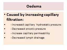
ETIOLOGY:
Edema
results from four causes:
I.
Increased hydrostatic
pressure in capillaries
and veins
due to chronic
CHF or
obstruction to venous return;
II.
Decreased
plasma
oncotic
pressure
III.
Increased
capillary permeability
in
endotoxemia
, part of the allergic
response,
vasculitis
and damage to the
vascular endothelium
;
or
IV.
Obstruction
to
lymphatic flow
Slide3Increased hydrostatic
pressure:
Symmetric ventral edema in
chronic
(congestive
) heart failure,
symmetric pulmonary
edema in acute
heart failure
Enzootic
calcinosis
of
cattle
udder edema in late
pregnancy
Local
edema by compressive
lesions on
veins (as in
thymic
lymphosarcoma
with compression
of the
cranial vena
cava)
draining
other anatomic locations
Local
edema in portal
hypertension due
to hepatic fibrosis causing
ascites
Slide4Decreased plasma
oncotic
pressure:
Decreased total protein concentration
in plasma
, and
particularly
decreased plasma
albumin concentration
, will
result in
symmetric ventral
edema
Hypoalbuminemia
can
result from
increased loss
(due to
bloodsucking parasites
or across the
gastrointestinal tract
, kidneys or into a
large third
space such as the pleural or
peritoneal cavities),
decreased production
(as
in chronic
hepatic failure) or
decreased intake
Slide5Increased capillary permeability:
Endotoxemia
Allergic edema as in
urticaria
and
angioneurotic
edema caused by
local liberation
of
vasodilators
Toxic damage to vascular
endothelium or
vasculitis
- in anthrax,
gas gangrene
and malignant edema
in ruminants
, edema disease of
pigs, mulberry
heart disease in pigs,
equine viral
arteritis
, equine
infectious anemia
,
purpura
hemorrhagica
in horses
, and
heartwater
(
cowdriosis
)
in ruminants
Slide6Obstruction to lymphatic
flow:
Part of the edema caused by
tumors or
inflammatory swellings
is lymphatic
obstruction
.
Extensive
fluid loss
also originates
from
granulomatous
lesions on
serous surfaces
.
Ascites
or hydrothorax
may result
PATHOGENESIS:
At the arteriolar end of
the capillaries
the hydrostatic pressure of
the blood
is sufficient to overcome its
oncotic
pressure
and fluid tends to pass into
the interstitial space
At the venous end
of the
capillaries the position is reversed
and fluid
tends to return to the
vascular system
Slide7CLINICAL
FINDINGS:
Anasarca
(
Accumulation of
edematous
transudate
in subcutaneous tissues)
Ascites
(Accumulation
of edematous
transudate
in peritoneal cavity)
Hydrothorax
Hydropericardium
S
wellings
are soft,
painless and
cool to the touch, and pit
on pressure
In
ascites
there is
distension
of the
abdomen
and the fluid can
be
detected
by a fluid thrill
on
tactile percussion,
fluid
sounds on
succussion
and
by
paracentesis
Slide8In the pleural cavities and pericardial sac
T
he
clinical signs i
nclude
restriction
of cardiac
movements, embarrassment
of respiration
and collapse of the
ventral parts
of the lungs
.
Muffled heart
sounds
and respiratory
sounds
are,
and
the presence
of fluid may be ascertained
by percussion
and
thoracocentesis
or
pericardiocentesis
More localized edemas cause
more localized
signs
:
P
ulmonary
edema
-respiratory distress
C
erebral edema-nervous signs
Thrombophlebitis
-
Head
edema due to
complete occlusion of
both jugular
veins
Slide9CLINICAL
PATHOLOGY:
Cytological
examination
for absence
of
inflammatory cells
Thoracocentesis
or
abdominocentesis
is useful to
differentiate the
causes of
fluid accumulation
Serum albumin
concentration and central
venous pressures
Hypoalbuminemia
-determine renal and
gastrointestinal systems and liver
Slide10TREATMENT:
Correcting
the
cause
Chronic (
congestive) heart
failure may need to be treated
with
digoxin
Hypoalbuminemia
may require the administration
of plasma
or plasma substitutes
Ancillary nonspecific
measures:
Restriction
of the amount of salt in the
diet
Use
of diuretics