
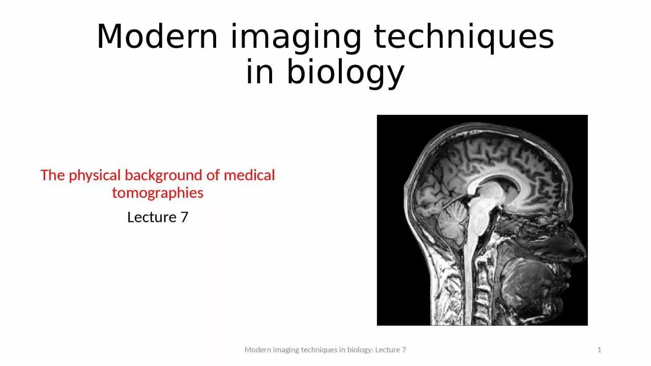
The physical background of medical tomographies Lecture 7 Modern imaging techniques in biology Lecture 7 1 MRI thematics Microscopic and macroscopic magnetization The Bloch equation T ID: 916023
Download Presentation The PPT/PDF document "Modern imaging techniques in biology" is the property of its rightful owner. Permission is granted to download and print the materials on this web site for personal, non-commercial use only, and to display it on your personal computer provided you do not modify the materials and that you retain all copyright notices contained in the materials. By downloading content from our website, you accept the terms of this agreement.


Slide1
Modern imaging techniques in biology
The physical background of medical tomographiesLecture 7
Modern imaging techniques in biology: Lecture 7
1
Slide2MRI thematics
Microscopic and macroscopic magnetization. The Bloch equation. T1 and T2
relaxation times. Magnetic resonance. 90° pulse. FID: free induction decay. Effective T
2.Pulse method for determining T2
and T
1
. Spin echo. Magnetic resonance spectroscopy.Imaging. Selective excitation and read out of a slice.Pulse sequences and contrast. Flow in MRI.fMRI. BOLD. Echo planar imaging (EPI). Spin echo (SE) and gradient echo (GE) for fast imaging.
Modern imaging techniques in biology: Lecture 7
2
https://mri.byu.edu
Slide3fMRI
pros and consAdvantages:non-invasivefair spatial and temporal resolution
can image the whole brain almost simultaneously Disadvantages:
measures indirect
signal
(
not the electric signal of neurons known to be
directly correlated with
information processing, i.e.,
brain function)measures the integral
signal
of
many neurons
Modern imaging techniques in biology: Lecture 7
3
Slide4Movie reconstruction from human brain activity
using fMRI
Modern imaging techniques in biology: Lecture 7
4
Scientists use brain imaging to reveal the movies in our
mind
http://news.berkeley.edu/2011/09/22/brain-movies/
Slide5T1
vs T2/T2* weigthed images
Normal
anatomical images of the
brain
are most often T1-weighted.fMRI images are
T2 or T
2* weigthed.
Modern imaging techniques in biology: Lecture 75
„
A third commonly used sequence is the
Fluid Attenuated Inversion Recovery (Flair)
. The Flair sequence is similar to a T2-weighted image except that the TE and TR times are very long. By doing so, abnormalities remain bright but normal CSF
(
c
erebrospinal
fluid)
is attenuated and made dark. This sequence is very sensitive to pathology and makes the differentiation between CSF and an abnormality much easier.
”
http://casemed.case.edu
Slide6fMRI:
functional MRISince 1890 we know
that hemodynamics
(blood flow, oxigenation)
strongly
correlates with neural activity. 1-5 secs after an increased
neuronal activiy
the blood flow increases ->
increased oxigenation, increased cerebral blood volume
(CBV)
Oxigen
transport: O
2 @red blood
cells (RBC) @Hemoglobin (Hb):
Modern imaging techniques in biology: Lecture 7
6
Hemoglobin (
Hb
),
Wikipedia
Slide7MRI can
measureTissue perfusionBlood
oxygenationBlood volume
Water diffusion
First
fMRI was aquired by a contrast agent to
measure local CBV (cerebral
blood volume). Then
CBV without contrast agent, and soon after
that
BOLD appeared.
Modern imaging techniques in biology: Lecture 7
7
http://www.neurologyindia.com
Slide8BOLD-contrast
: Blood oxigenation level dependent contrast
Fe in
the Heme group
is in
high
spin state and thus paramagnetic in deoxy hemoglobin deoxy-Hb. 4 unpaired
electrons.http://mriquestions.com/bold-contrast.html
Modern imaging techniques in biology: Lecture 7
8
Heme
group
:
iron (Fe) ion held in a heterocyclic ring, known as a porphyrin
BOLD
since
1990
:
hemodynamic
response
.
Slide9Origin of BOLD
contrastModern imaging techniques in biology: Lecture 79
Paramagnetic
deoxyhemoglobin
(D) confined to red blood cells causes a local field distortion in and around the vessel.
„
The
presence of paramagnetic
deoxyhemoglobin
within red blood cells creates local magnetic field distortions (susceptibility gradients) in and around blood vessels. These local field disturbances cause nearby stationary and slowly moving spins to have different resonance frequencies and phase shifts. The resultant intravoxel
dephasing
is a classic T2*-shortening effect most prominent near larger veins and accentuated by use of GRE sequences with echo times (
TEs
) close to T2*. The effect scales linearly with field strength (
Bo
) and is the dominant mechanism for BOLD contrast at 1.5T
.
”
http://mriquestions.com/bold-contrast.html
Slide10BOLD contrast
: T2 and T2*
Whereas m
agnetic susceptibility of the
d
iamagnetic oxy-Hb is similar to the susceptibility of tissues,
paramagnetic deoxy-Hb
is different. The presence of
deoxy-Hb results in magnetic field inhomogeneity.
w
here
b
z
is
the
local
fluctuating
magnetic
field
.
where
is
the
static
field
inhomogeneity
.
Both
T
2
and T
2
*
will
correlate
strongly
with
the
BOLD
signal
.
Modern imaging techniques in biology: Lecture 7
10
Slide11Echo-planar
imaging (EPI): MRI in a fraction of a second
1 excitation per image. A single
RF shot can
scan
the entire k space.
,
Modern imaging techniques in biology: Lecture 7
11
Slide12Echo-planar imaging
(EPI)Modern imaging techniques in biology: Lecture 7
12
M. K.
Stehling
et al. Science 254, 43-50 (1991).
Ways to scan
the k space
or Fourier space. A single RF pulse
can
scan the
entire k space
.
Slide13Gradient
echo (GE) versus spin echo (SE)The SE
pulse sequence has a 90° excitation pulse (GE has a small excitation pulse), and SE refocuses some of the dephasing which occurs during the echo time using a 180° refocusing RF pulse.
Because only one RF pulse is applied in GE, the echo can be recorded more quickly, resulting in a shorter echo time. If low flip angles are used, TR can also be shorter.
Thus
GE is preferred for rapid imaging techniques. GE image contrast is dictated by T2*, unlike in SE where image contrast is dictated by T2. In SE the signal-to-noise ratio is higher.http://www.revisemri.com
Modern imaging techniques in biology: Lecture 7
13
Spin-
echo
(SE) versus
gradient
echo
(GE)