
httpilivesciencecomimagesi000024292iFFneurons120208jpg1328727600 What is the nervous system The nervous system is a complex collection of nerves and specialized cells known as neurons that transmit signals between different parts of the body It is essentially the bodys electric ID: 1047595
Download Presentation The PPT/PDF document "Nervous System By: Jenny Jin" is the property of its rightful owner. Permission is granted to download and print the materials on this web site for personal, non-commercial use only, and to display it on your personal computer provided you do not modify the materials and that you retain all copyright notices contained in the materials. By downloading content from our website, you accept the terms of this agreement.


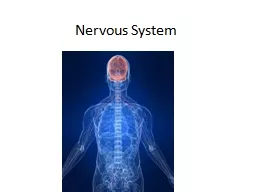
1. Nervous SystemBy: Jenny Jinhttp://i.livescience.com/images/i/000/024/292/iFF/neurons-120208.jpg?1328727600
2. What is the nervous system?The nervous system is a complex collection of nerves and specialized cells known as neurons that transmit signals between different parts of the body. It is essentially the body’s electrical wiring. The functions of the nervous system can be separated into two subdivisions: somatic (voluntary) and autonomic (involuntary).
3. Somatic Nervous SystemNerves that connect the brain and spinal cord with muscles and sensory receptors in the brain; this system enables our voluntary control of muscles, as well as our reception of sights, sounds, sensations, tastes and smells.http://studydroid.com/imageCards/03/ki/card-3820362-back.jpg
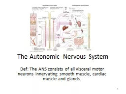
4. Autonomic Nervous SystemRegulates certain body processes that work without conscious effort; such as digestion, salivation, heart rate, perspiration and the “fight or flight” response.http://www.chiro1source.com/assets/images/autonomic_nervous_lateral_labeled_mediumb_prod.jpg
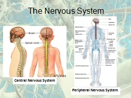
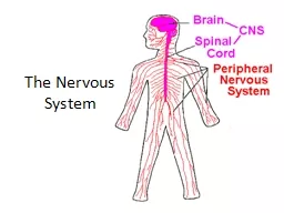
5. Central Nervous SystemThe central nervous system is that part of the nervous system that consists of the brain and spinal cord.Major parts:BrainSpinal cordPeripheral Nervous System The peripheral nervous system connects the central nervous system to sensory organs (such as the eye and ear), other organs of the body, muscles, blood vessels and glands. Major parts:Nerve fibers that connect the spinal cord to all parts of the bodyThe brain sends messages through the spinal cord and nerves of the peripheral nervous system to control the movement of the muscles and the function of internal organs.
6. Interaction of Two NeuronsBy: Jenny Jin
7. Simple Reflex ArcBy: Jenny JinA reflex arc defines the pathway by which a reflex travels, from the stimulus to sensory neuron to motor neuron to reflex muscle movement.Reflexes are involuntary, instantaneous movements in response to a stimulus.Reflex arcs involve three neurons: stimulus, interneuron, and motor neuron. The stimulus (e.g., needle stick, hot surface) stimulates the pain receptors of the skin, which initiates an impulse in the sensory neurons.This impulse travels to the spinal cord, where the sensory neuron makes a synapse with the interneuron. The impulse then travels to one or more motor neurons that transmit the impulse to the muscles through the effectors, causing them to contract and pull away from the stimulus.Reflexes do not require involvement of the brain; this is how reflexes can be so instantaneous.
8. Cerebral HemispheresLeft Cerebral HemisphereRight Cerebral HemisphereThe right cerebral hemisphere controls movement of the left side of the body. In most people, areas that govern spatial perceptions reside in the right hemisphere.By: Jenny JinThe left cerebral hemisphere controls movement of the right side of the body.In most people, the areas that control speech are located in the left hemisphere.The brain is separated into the frontal, occipital, and parietal lobes.The frontal lobe is associated with executive functions and motor performance.The occipital lobe is the visual-processing center of the brain.The parietal lobe is associated with sensory skills.
9. DiencephalonBy: Jenny JinMain structures of the diencephalon include the hypothalamus, thalamus, and the epithalamus (including the pineal gland). The diencephalon relays sensory information between brain regions and controls many autonomic functions of the peripheral nervous system.Hypothalamus: maintains homeostasisThalamus: controls sensory perception, movement, sleep, and consciousnessEpithalamus: produces important hormones
10. Brain stemBy: Jenny JinThe brain stem is one of the most basic regions of the human brain, yet it is one of the most vital regions for our body’s survival. It forms the connection between the brain and the spinal cord, maintains vital control of the heart and lungs, and coordinates many important reflexes.Midbrain: a vital aspect of our neural 'information superhighway,' which transfers visual and auditory input to the brain and motor information from the brainPons: responsible for feeling in the face, controls the muscles that are responsible for biting, chewing, and swallowing, allows the eyes to look from side to side, controls facial expressions, allows sound to move from the ear to the brainMedulla: helps regulate breathing, heart and blood vessel function, digestion, sneezing, and swallowing. This part of the brain is a center for respiration and circulation.
11. CerebellumThe cerebellum modifies the motor commands of the descending pathways to make movements more adaptive and accurate.Maintains balance and postureCoordinates voluntary movementsFacilitates motor learning and cognitive functions through trial-and-error
12. How a Nerve Impulse TravelsWhen a neuron is at rest, it is in a state of polarization and contains membrane potential. There is an excess of sodium (Na+) ions outside of the cell membrane that create a positive charge. Similarly, there is an excess of potassium (K+) ions inside the cell along with negatively charged molecules that produce a negative charge inside the cell membrane. This is the cell’s resting potential. When a neuron is stimulated, either from direct sensory input or another neuron, ion channels in the cell membrane open, and sodium ions rush in. The inside of the cell becomes positively charged, the cell goes through a depolarization to its threshold, and an action potential is created that transmits the stimulus down the axon.At the end of the axon, the nerve impulse reaches the synapse. Neurotransmitters are then released to carry the nerve impulse across the synapse to the dendrites of the next neuron.The neuron is then in a refractory period before receiving the next message.
13. http://www.moleculardevices.com/sites/default/files/flipr-assay-membranepot-principle_Figure_450.jpghttps://upload.wikimedia.org/wikipedia/commons/f/fb/Basis_of_Membrane_Potential2.pnghttp://www.helcohi.com/sse/images/body/1-4ci.gifhttp://www.biologymad.com/nervoussystem/propagation2.jpg
14. NeurotransmittersNeurotransmitters are the brain chemicals that communicate information throughout our brain and body. They relay signals between neurons. Neurotransmitters are released by axons into the fluid of the synapse. Some of these chemicals bind to receptor sites on the corresponding dendrite, some of them return to the axon, and some of them are broken down, or metabolized.Neurotransmitters travel in vesicles down the axon; the vesicles then fuse with the synapse terminals and the neurotransmitters are released.Depending on which kind of neurotransmitter (IPSP vs. EPSP), the postsynaptic cell can either become more (EPSP) or less (IPSP) likely to fire an action potential. This action potential will lead to a continuation of the message.
15. IPSP vs. EPSPMany different molecules can act as neurotransmitters.When small amounts of neurotransmitters are released, the effect on the cell’s membrane potential varies in proportion to the amount of neurotransmitter released.IPSPs are neurotransmitters that are inhibitory; they open a set of ion channels that allow negatively charged ions to enter the cell.EPSPs are neurotransmitters that are excitatory; they cause the inside of the cell to become more positive than the outside.The IPSPs and EPSPs determine the graded potential; they can cancel out each other in a cell.EPSPs stimulate the brain, while IPSPs calm the brain and help create balance.
16. Amyotrophic Lateral SclerosisWhat is ALS?A rapidly progressive, invariably fatal neurological disease that attacks the neurons that control voluntary muscles (somatic nervous system)Both the upper motor neurons and the lower motor neurons degenerate or die, and stop sending messages to muscles. Unable to function, the muscles gradually weaken, waste away, and have very fine twitches. Eventually, the ability of the brain to start and control voluntary movement is lost.Signs and SymptomsTwitches, cramps, tight muscles, muscle weakness in an arm or leg, slurred and nasal speech, difficulty chewing or swallowingWhen symptoms begin in the arms or legs, it is known as “limb onset” ALS. If they begin in speech, it is known as “bulbar onset” ALS.Muscle weakness will then spread to other parts of the body; eventually, people with ALS will not be able to stand, walk, use their hands, and chew and swallow. They will also lose the ability to breathe on their own.http://static1.squarespace.com/static/52ce318fe4b05da9bada2a07/t/55404136e4b06d7f0e88dc69/1430274359602/http://ichef.bbci.co.uk/news/624/media/images/77299000/jpg/_77299767_hawking.jpgStephen Hawking
17. Amyotrophic Lateral Sclerosis (cont.)PrevalenceMore than 12,000 people in the U.S. have ALS, which is about 3.9 people per every 100,000. ALS is one of the most common disorders of the nervous system; men are affected more than womenIn most cases, ALS appears randomly and usually shows up in people who do not have a familial history of ALS.Treatment OptionsThere is not yet a cure for ALS; however, a drug named Riluzole was developed in 1995 which expanded lifespan by a few monthsPhysicians and therapists provide medical and physical therapy, as well as special equipment to help make patients as mobile and comfortable as possibleFeeding tubes and mechanical ventilation may assist the eventual degeneration of the mouth and the lungsEmotional and mental therapy is also vital; though their muscles are atrophying, the patients’ brains are well-functioning, which can lead to anxiety and depression, as the patients are aware of their inevitable loss of functionhttp://www.neurology.org/content/52/3/504/F2.large.jpg
18. EpilepsyWhat is epilepsy?Epilepsy is a central nervous system disorder (neurological disorder) in which neuron activity in the brain becomes disrupted, causing seizures or periods of unusual behavior, sensations and sometimes loss of consciousness. Epilepsy is categorized for two factors: idiopathic or symptomatic, and generalized or partialSigns and SymptomsMost apparent symptom is the appearance of seizuresMay cry out, stiffen for several seconds, and then make rhythmic movements of the arms and legsMay appear to not be breathing and may turn blueTemporary confusionA staring spellUncontrollable jerking movements of the arms and legsLoss of consciousness or awarenessPsychic symptomsGeneralizedPartialIdiopathicNo apparent cause; involves the whole brainNo apparent cause; involves one area of the brainSymptomaticCause is identified; involves the whole brainCause is identified; involves one area of the brain
19. Epilepsy (cont.)PrevalenceEpilepsy is a relatively common condition, affecting 0.5% to 1% of the population. In the United States, about 2.5 million people have epilepsy and about 9% of Americans will have at least one seizure in their lifetimes.Treatment OptionsDoctors generally begin by treating epilepsy with medication.At least half of all people newly diagnosed with epilepsy will become seizure-free with their first medication. If anti-epileptic medications don't provide satisfactory results, doctors may suggest surgery or other therapies.In surgery, your doctor removes the area of your brain that's causing the seizures.A ketogenic diet, one that is high fats and low in carbohydrates, may help reduce incidence of seizures.http://seizures.dolyan.com/wp-content/uploads/2011/10/Handling-People-Experiencing-Grand-Mal-Seizures.jpg
20. Bibliographyhttp://www.livescience.com/22665-nervous-system.htmlhttp://www.medicinenet.com/script/main/art.asp?articlekey=2667 https://www.nichd.nih.gov/health/topics/neuro/conditioninfo/Pages/parts.aspxhttp://www.ninds.nih.gov/disorders/amyotrophiclateralsclerosis/detail_ALS.htmhttps://www.boundless.com/physiology/textbooks/boundless-anatomy-and-physiology-textbook/the-peripheral-nervous-system-pns-13/reflexes-136/components-of-a-reflex-arc-727-2295/ http://www.webmd.com/epilepsy/http://www.mayoclinic.org/diseases-conditions/epilepsy/home/ovc-20117206http://courses.washington.edu/psych333/handouts/coursepack/ch05-Signalling_in_neurons.pdfhttps://www.neurogistics.com/TheScience/WhatareNeurotransmi09CE.asphttp://nawrot.psych.ndsu.nodak.edu/courses/465projects08/schizophrenia/neurotransmitters.htmhttps://faculty.washington.edu/chudler/synapse.htmlhttps://www.nlm.nih.gov/medlineplus/ency/imagepages/18011.htmhttps://www.boundless.com/psychology/textbooks/boundless-psychology-textbook/biological-foundations-of-psychology-3/structure-and-function-of-the-brain-35/cerebral-hemispheres-and-lobes-of-the-brain-153-12688/ http://biology.about.com/od/anatomy/p/diencephalon.htm http://www.innerbody.com/image_nerv01/nerv46.htmlhttp://neuroscience.uth.tmc.edu/s3/chapter05.htmlCampbell Biology in Focus - AP Edition (High School), 1e by Urry/Cain/Wasserman/Minorsky/Jackson/Reece