
Patternn dystrophies PD are a group of autosomal dominant diseases caused by mutations in the RDS gene They are characterized by lipofuscin accumulation in the retinal pigment epithelium RPE The diagnosis of PD is based on the pattern of pigment deposition in the RPE ID: 928472
Download Presentation The PPT/PDF document "Butterfly-shaped pattern dystrophy: a ca..." is the property of its rightful owner. Permission is granted to download and print the materials on this web site for personal, non-commercial use only, and to display it on your personal computer provided you do not modify the materials and that you retain all copyright notices contained in the materials. By downloading content from our website, you accept the terms of this agreement.


Slide1
Butterfly-shaped pattern dystrophy: a case report
Patternn
dystrophies (PD) are a group of
autosomal dominant diseases, caused by mutations in the RDS gene. They are characterized by lipofuscin accumulation in the retinal pigment epithelium (RPE). The diagnosis of PD is based on the pattern of pigment deposition in the RPE. Metamorphopsia and a slight decrease in vision are common presenting symptoms, although lots of cases are just asymptomatic. According to the distribution of the pigment, PD are separated into 5 different categories. Butterfly-shaped pattern dystrophy (BPD) is one of them, and is defined by a bilateral accumulation of yellowish or pigmented material in a butterfly-like pattern at the level of the RPE.
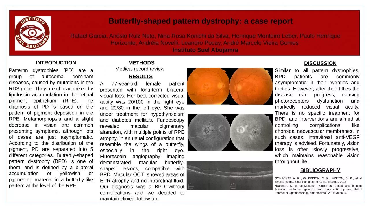
A 77-year-old female patient presented with long-term bilateral visual loss. Her best corrected visual acuity was 20/100 in the right eye and 20/80 in the left eye. She was under treatment for hypothyroidism and diabetes mellitus. Fundoscopy revealed macular pigmentary alteration, with multiple points of RPE atrophy, in an usual configuration that resemble the wings of a butterfly, especially in the right eye. Fluorescein angiography imaging demonstrated macular butterfly-shaped lesions, compatible with BPD. Macular OCT showed areas of EPR atrophy and no intraretinal fluid. Our diagnosis was a BPD without complications and we decided to maintain clinical follow-up.
INTRODUCTION
.
METHODS
RESULTS
DISCUSSION
Similar to all pattern dystrophies, BPD patients are commonly asymptomatic in their twenties and thirties. However, after their fifties the disease can progress, causing photoreceptors dysfunction and markedly reduced visual acuity. There is no specific treatment for BPD, and interventions are aimed at controlling complications like choroidal neovascular membranes. In such cases, intravitreal anti-VEGF therapy is advised. Fortunately, vision loss is often slowly progressive, which maintains reasonable vision throughout life.
BIBLIOGRAPHY
SCHACHAT, A. P. , WILKINSON, C. P., HINTON, D. R., et al; Ryan's Retina. 6 ed. Rio de Janeiro: Ed. Elsevier, 2017Rahman, N. et. al. Macular dystrophies: clinical and imaging features, molecular genetics and therapeutic options. British Journal of Ophthalmology, bjophthalmol–2019–315086.
Rafael Garcia, Anésio Ruiz Neto, Nina Rosa Konichi da Silva, Henrique Monteiro Leber, Paulo Henrique Horizonte, Andréia Novelli, Leandro Pocay, André Marcelo Vieira Gomes Instituto Suel Abujamra
Medical record review