
Benign cephalic hix00730074iocytosis BCH is a rare benign selfhealing nonLangerhans hix00730074iocytosis that presents in infancy and early childhood Gianotti et al x00660069rx007300 ID: 939668
Download Pdf The PPT/PDF document "Hix00730074iocytoses are a diverse group..." is the property of its rightful owner. Permission is granted to download and print the materials on this web site for personal, non-commercial use only, and to display it on your personal computer provided you do not modify the materials and that you retain all copyright notices contained in the materials. By downloading content from our website, you accept the terms of this agreement.


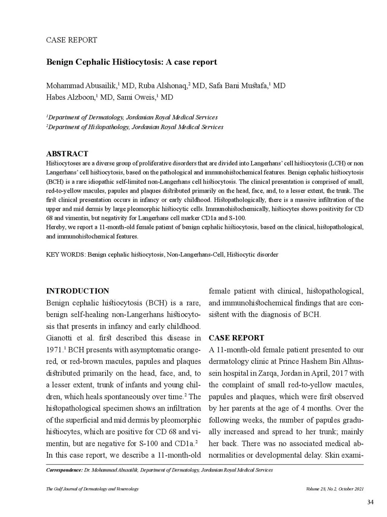
Hi�iocytoses are a diverse group of proliferative disorders that are divided into Langerhans’ cell hi�iocytosis (LCH) or non Langerhans’ cell hi�iocytosis, based on the pathological and immunohi�ochemical features. Benign cephalic hi�iocytosis Benign cephalic hi�iocytosis (BCH) is a rare, benign self-healing non-Langerhans hi�iocytosis that presents in infancy and early childhood. Gianotti et al. �r� described this disease in red, or red-brown macules, papules and plaques Volume 28, No.2, October 2021The Gulf Journal of Dermatology and Venereology nation at the time of presentation showed multiple, scattered red-to-yellow macules, papules and plaques that were 2 to 8 mm over the face Di�use cell in�ltrates were observed throughout the High power view demon�rating proliferation of large, epithelioid hi�iocytic cells with eosinophilic cy A Laboratory inve�igation showed normal hematologic and biochemical indices. To rule out emia, lipid pro�le (chole�erol, triglycerides, HDL, LDL) and serum protein electrophoresis (IgA, IgG and IgM) were done and found to be normal. Abdominal ultrasound was normal without organomegally.Hi�ological examination of the biopsy specimen taken from a lesion on the back showed a normal epidermis and massive in�ltration of the super�cial and mid dermis by large, pleomorphic epitheliod hi�iocytic cells with abundant eosinophilic cytoplasm, hyperchromatic nuclei and large nucleoli. Mitotic �gures, cytoplasmic lipids and multinucleated giant cells were absent. There was a
n inter�itial in�ammatory in�ltrate composed of moderate numbers of lymphocytes and scattered eosinophils (Fig. 2). Immunohi�ochemi�ry showed that the cells �ained positive for CD 68 and vimentin, but negative for the S100 protein and CD1a (Fig. 3). According to the clinicopathologic �ndings, the disease was diagnosed as benign cephalic hi�iocytosis.gression of the whole lesions from the face and Mohammad Abusailik Multiple, scattered yellow-red macules, papules and A B Benign Cephalic Hi�iocytosis: A case reportperpigmented macules (Fig. 4). No scarring has observed. Skin biopsy was not taken at this time due to patient’s parent hindrance. A B D Fig. 3Immunohi�ochemical �aining shows positive for CD Vimentin, negative for (C)BCH is a rare non-Langerhans cell hi�iocytic sented as a self-healing asymptomatic papules and plaques a�ecting primarily the head and The lesions are 1 to 8 mm in diameter and its colors range from yellow to red brown. The lesions �r� appear on the face and neck and subsequently extend to the trunk and rarely to buttock and upper and lower extremity. Consistent with other reported cases, our patient had multiple, 2 to 8 mm yellow-red macules, papules and plaques that �r� appeared on the face and later spread to the back. There is no involvement of mucous membranes, palmoplantar skin, and internal organ in this disease. The �r� presentaFig. 4 complete regression of the lesions with resultant hyper A B tion of the lesions ranges from 3 to 36 months In approximately half of cases, the patien
ts are younger than 6 months. There is no gender predilection with males and females are equally a�ected. Complete regression of the lesions occurs within 50 months, on average. The papules �atten over time, eventu In our case, complete resolution of the face and back lesions occurred within 32 months, with some lesions developed hyperpigmented macules.normalities or sy�emic involvement. However, tes mellitus were reported in association with BCH. Diabetes insipidus results of in�ltration of the pituitary gland by hi�iocytic cells, this �nding has been noticed more commonly in xanthoma disseminatum and Langerhans cell hi�io Furthermore, there is a report of BCH progressing intojuvenile xanthogranuloma under the in�uence of a Varicella-zo�er infec Therefore, careful regular examination is recommended for all patients with BCH, to monitor for progression, exacerbation, or internal organ involvement. Apart from this careful examination, no treatment or other intervention are required, considering that the lesions will heal spontaneously, although topical 1% rapamycin has been used successfully to treat facial lesions The hi�opathologic hallmark of BCH is a well-circumscribed su�cient in�ltration of the upper and mid dermis by hi�iocytic cells. There is no epidermotropism. There is a mixed in�ammatory dermal in�ltrate composed of lymphocytes and rare eosinophils. Hi�iocytes are typically large, pleomorphic, with abundant eosinophilic cytoplasm and oval, hyperchromic, vesicular and sometimes indented nuclei, often with very prominent large nucleoli. Mitos
es are absent. Immunohi�ochemically, Langerhans cell markers, CD1a and S-100, are negative. Whereas histiocytic markers, CD68, are positive. All these hi�opathological �nding were observed in the hi�ological specimen of our case.BCH mu� primarily be di�erentiated from small nodular form of juvenile xanthogranuloma (JXG) and generalized eruptive hi�iocytoma Other di�erential diagnoses include LCH, verruca vulgaris, urticaria pigmentosa, sarcoidosis and multiple Spitz nevi. Hi�ologic examination can di�inguish Lesions of BCH tosa. The lesions of small nodular form of JXG are more widely di�ributed over the entire skin with higher incidence of extracutaneous lesions especially ocular involvement. Hi�opathological examination of JXG reveals the characteristic Touton giant cells and can easily di�eren GEH is observed more commonly in adults, and presents with a more widespread axial di�ribution of recurrent crops sal surfaces involvement. The hi�opathological �nding may be similar to BCH. LCH has more widespread skin and internal organ involvement. Immunohi�ochemically, they are positive for S100 and CD1a. Ultra�ructurally, intracytoplasGianotti F, Caputo R, Ermacora E. Singular “infantile hi�iocytosis with cells with intracytoplasmic vermiform particles”. Bull Soc Fr Dermatol Syphiligr. 1971; 78:232-33.Mohammad Abusailik Benign Cephalic Hi�iocytosis: A case reportKim BC, Choi WJ, Seung NR, et al. A Case of Benign Cephalic Hi�iocytosis.Ann Dermatol. 2011;23(Suppl 1):S16-S19.Koca R, Bektaş S, Al
tinyazar HC, Sezer T. Benign Cephalic Hi�iocytosis: A Case Report.Ann Dermatol. 2011; 23(4):508-511. ules in a child. Benign cephalic hi�iocytosis. Arch Polat Ekinci A, Buyukbabani N, Baykal C. Novel Clinical Observations on Benign Cephalic Hi�iocytosis in a Large Series. Pediatr Dermatol. 2017; 34(4):392-397. Jih DM, Salcedo SL, Jaworsky C. Benign cephalic hi�iocytosis:a case report and review. J Am Acad We�on WL, Travers SH, Mierau GW, Heasley D, Fitzpatrick J. Benign cephalic hi�iocytosis with dia�er C, Orozco-Covarrubias L, Ruiz-Maldonado R. Benign cephalic hi�iocytosis preceding the development of insulin-dependent diabetes mellitus. Pediatr Rodriguez-Jurado, Rodolfo M.D.; Duran-McKin�er, Benign Cephalic Hi�iocytosis Progressing Into Juvenile Xanthogranuloma: A Non-Langerhans Cell Hi�iocytosis Transforming Under the In�uence of a Virus?, Patsatsi A, Kyriakou A, Sotiriadis D. Benign cephalic hi�iocytosis: case report and review of the literature. Pediatr Dermatol. 2014; 31(5):547-50. 11.Habeshian K, Silverman RA, DeKlotz CMC. Treatment of benign cephalic hi�iocytosis with topical 1% rapamycin ointment. Pediatr Dermatol. 2019; 36(3):411-413.ules in a child. Benign cephalic hi�iocytosis. Arch Dermatol. 1995; 131:610-11, 613-14.seminated juvenile xanthogranulomatosis benign cephalic hi�iocytosis? Pediatr Dermatol. 2005; 22:40-43.Caputo R, Ermacora E, Gelmetti C, Berti E, Gianni E, Nigro A. Generalized eruptive hi�iocytoma in children. J Am Acad Dermatol. 1987; 17:449-54. Volume 28, No.2, October 2021The Gulf Journal of Dermatology and Venereo