

DIAGNOSIS rapid inflammation middle ear effusion MEE MEE without acute inflammation AOM OME inflamation Signs ID: 752758
Download Presentation The PPT/PDF document "Acute otitise media &OTITIS MEDIA ..." is the property of its rightful owner. Permission is granted to download and print the materials on this web site for personal, non-commercial use only, and to display it on your personal computer provided you do not modify the materials and that you retain all copyright notices contained in the materials. By downloading content from our website, you accept the terms of this agreement.


Slide1
Acute otitise media
&OTITIS MEDIA WITH EFFUSIONSlide2
DIAGNOSISrapid inflammation + middle ear effusion (MEE)
MEE
without
acute inflammation
AOM
OME:
inflamation
Signs:
bulging
or
fullness
or
erythema
or
perforation of the TM with
otorrhea
Symptoms:
otalgia
, irritability, and fever Slide3
EPIDEMIOLOGY (
AOM) is the most frequent diagnosis in sick children
in US
approximately $5 billion in US
otitis
media
39
%
of children
by
9 months
and
62%
of children
by 2
years
of age
occurs
in older children,
adolescents,and
adults
.
peak
incidence
of
AOM was during
the first 6 to 12 months
of
life
OME
is asymptomatic. approximately 65% of OME episodes in children 2 to 7 years of age resolve within 1 month
.
difficult to determine the “true” incidence of OME Slide4
PHYSICAL EXAMINATION Ears Head and
neck
C
raniofacial
anomalies
(
Down and
Treacher
Collins
)
O
ropharynx
(
bifid uvula or
cleft
palate)
Hypernasality
(
velopharyngeal
insufficiency)
H
yponasality
(
obstructing
adenoids or nasal obstruction due to
nasal polyposis
or deviated
septum)Slide5
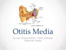
Pneumatic otoscopy
M
iddle ear
TM and its mobility.
normal TM : translucent
concave
moves
with
positive and negative pressure
.
landmark:
handle (manubrium) of the
malleus
. umbo:
in the center of the TM.
Note:
position, color, degree of translucency
,
mobilitySlide6Slide7
position position of the tympanic membrane is the most critical characteristic in distinguishing AOM from OME normal position is neutral negative
pressure: retracted TM
fullness (infection)
bulging:large
amount of infected fluid (
posterosuperior
area) when bulging: the malleus is obscured Slide8
Translucency normal TM is translucent with fluid: cloudy or opaque Air
fluid levels are more suggestive of OME than
AOMSlide9
color “red” TM that is full or bulging often is a sign of
AOM
A
pink
,
gray, yellow
, or
blue
retracted TM with reduced or no
mobility
usually is seen with
OME
.
red but translucent
TM is a typical finding in a
crying or sneezing
infant
, Slide10Slide11Slide12Slide13
TYMPANOMETRY inconclusive otoscopy difficult
otoscopy
children older than 6 months Slide14
tympanometry −400 to +200 daPa(decapascals
).
flat
or round pattern(TW>350
daPa
)with a small ear canal volume:
MEE
flat pattern with a large ear canal volume :
perforation
or
a patent
tympanostomy
tube.
normal middle ear: peak pressure 0
daPa
no OME : TW<150
daPa
OME: TW> 350
daPa
TW=150-350
daPa
presence or absence of OME is determined by
otoscop
ySlide15
AUDIOMETRYMEE usually results in a mild to moderate conductive hearing loss and causes delay in speech and language development
Slide16
OAEcochlear function (outer hair cells) -newborn hearing screening :fast and easy
MEE
may confound the results
.
ABRSlide17
PATHOPHYSIOLOGY ANDPATHOGENESIS
multifactorial
with various overlapping factors
1.infection(bacteria,viral
)2.Host factors(Allergy,immunology,gender,race,age,gentic
)
3.anatomic/physiologic(
eustachian
tube,cleft
palat
)
4.Enviroment factor(
daycar,tobacco
smoke exposure seasonality breast/bottle
feeding,pacifier,obisitySlide18
EUSTACHIAN TUBE FUNCTIONThe eustachian tube in the infant is shorter, wider, and more horizontal By the age of 7
years prevalence of
otitis media is low
.
Slide19
INFECTION in AOM
Streptococcus
pneumoniae
most common
Haemophilus
influenzae
Moraxella
catarrhalis
Streptococcus
pyogenes
other miscellaneous bacteria
in chronic OME
,
H.
influenzae
most common pathogen
S.
pneumoniae
M.
catarrhalis
other bacteria Slide20
Viruses respiratory syncytial
virus (RSV
)
influenzavirus
adenoviruse
parainfluenza
virus rhinoviruses Slide21
ALLERGY AND IMMUNOLOGY mechanism is not
understood,it
may be:
(1)
the middle ear is a “shock organ
” (
target
)
(2
)
induce inflammatory swelling of
the
eustachian
tube
mucosa
(3
) inflammatory
obstruction
of the
nose
(4)
bacteria-laden allergic
nasopharyngeal secretions
may be aspirated into the
midle
earSlide22
RISK FACTORESSlide23
Host-Related Factors
Age
.
highest incidence
6 -
11 months
of
age,
first episode <
6
or 12 months
a
powerful
predictor
of recurrence.
first episode of MEE <
2 months
is higher
risk for persistent fluid during their
first year
of
life
Sex
.
no difference between male & female
Prematurity
controversy
Allergy
.
controversy .
Immunocompetence
.
HIV
demonstrate a significantly higher recurrence
Slide24
Cleft Palate/Craniofacial Abnormality. Infants < 2 year
with
unrepaired cleft
palate Surgical
repair reduces otitis
media Anatomic
or functional eustachian tube abnormalities
Down
syndrome:
low resistance of the
tube
poor active
eustachian
tube
reflux of nasal secretions into the middle
ear.Slide25
Environmental FactorsUpper Respiratory Infection/Seasonality Rhinovirus, RSV,adenovirus
, and coronavirus
Day Care/Home
care
day-care centers more
tympanostomy tubes inserted than home care
Tobacco
Smoke Exposure
passive exposure to smoking
Breastfeeding/Bottle Feeding
Pacifier
Use
unclear.
ObesitySlide26
SYMPTOMATIC THERAPY ibuprofen 10 mg/kg
Auralgan
® (combination of
antipyrine
,
benzocaine , and glycerin
)
topical aqueous
lidocaine
(lignocaine) ear drops
topical herbal extract
Otikon
Otic
solution
Decongestants and antihistamines:
no
benefit
potential for
delayed resolution of middle ear
fluid
increased medication
side effectsSlide27
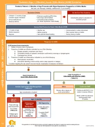
ANTIBIOTIC THERAPY VERSUS OBSERVATION < six
months
antibacerial
therapy regardless of degree of diagnostic certainly
six months to two years
,
antibacterial therapy
is
when: certain diagnosis of AOM uncertain diagnosis
but the illness is severe
(moderate
to severe
otalgia
or fever ≥39ºC in the previous 24 hours
).
Observation
when
diagnosis is not certain
and
illness is not severe.
>
two years
,
antibacterial therapy
when: certain diagnosis
and
illness is
severe
Observation
when:
certain diagnosis
but
illness is not
severe
uncertain diagnosis. Slide28
ANTIMICROBIAL THERAPY Seventeen antimicrobial drugs (16 oral and 1 parenteral preparation) two otic preparations (eg
,
ofloxacin
otic
and ciprofloxacin-dexamethasone otic)
for treatment of AOM with otorrhea in children with
tympanostomy
tubes in place or tympanic membrane perforation Slide29
Antimicrobial agents available for treatment of acute otitis media
Most used drugs
Others
Amoxicillin
Cephalexin
Amoxicillin-
clavulanate
*
Cefaclor
Cefuroxime
axetil
*
Loracarbef
Ceftriaxone IM or IV*
Cefixime
Erythromycin +
sulfisoxazole
•
Ceftibuten
Azithromycin
•
Cefprozil
Clarithromycin
•
Cefpodoxime
Trimethoprim-
sulfamethoxazole
•
Δ
Cefdinir
Ofloxacin
otic
◊
Trimethoprim
Ciprofloxacin-dexamethasone
otic
◊ Slide30
First-line therapy amoxicillin of 80 to 90 mg/kg per day maximum dose of 3 g/day Amoxicillin-clavulunate
AOM by
an amoxicillin-resistant
otopathogen:
antibiotictherapy
in the previous 30 days, particularly beta-lactam antibiotics concurrent purulent conjunctivitis (otitis-conjunctivitis syndrome usually is caused by
nontypeable
H.
influenzae
, which is frequently resistant to beta-lactam antibiotics)
receiving
amoxicillin
for chemoprophylaxis of recurrent AOM (or urinary tract infection) Slide31
Penicillin allergy Non-type 1 reactions : Cefdinir 14 mg/kg per day
Cefpodoxime
10 mg/kg per day once
daily
Cefuroxime – cefuroxime axetil suspension:
A
single intramuscular dose of
ceftriaxone
50 mg/kg
If clinical signs persist, a second dose is administered and, if necessary, a third dose
.
Type 1 reactions
:
azithromycin
, and
clarithromycin
.
Trimethoprim-
sulfamethoxazoleSlide32
Duration of therapy < 2 years old : 10 days >2 years old:
5-7 days single
dose of
azithromycin
has been approved by the FDA Slide33
Treatment failure Lack of improvement by 48 to 72 hours : another disease is present
the initial therapy was not adequate
.
Inadequate therapy :
organism resistant to beta-lactam antibiotics
Persistent
MEE
after the resolution of acute symptoms
is not an indication of treatment failure
or an indication for additional antibiotic
therapy
high-dose
amoxicillin-
clavulanate
90 mg/kg per day amoxicillin and 6.4 mg/kg per day of
clavulanate
Tympanocentesis
for
patients with persistently refractory AOM,
to define the
etiology Alternatively
,
use of
levofloxacin
and/or
tympanostomy
tube placement may be appropriate
. Slide34
Recurrent AOM signs and symptoms of AOM (fever, pain, bulging tympanic membrane) soon after completion of successful treatment.(within 30 days)
bulging of the tympanic membrane
and signs of inflammation.
p
ersistent
MEE in a child with a febrile upper respiratory infection may be misinterpreted as a recurrent episode.
Parenteral
ceftriaxone
50 mg/kg per day for 3 days or possibly every 36
hour
levofloxacin
10
mg/kg every 12
hrs
recurrence
more than 30 days
is most often due to a different
pathogene
: high
dose
amoxicillin-
clavulanate
Tympanostomy
tube insertion may be warranted for
children with recurrent AOMSlide35
Tympanic membrane perforation acute otorrhea, 10 days of oral therapy
topical
therapy
for the well-appearing, immunocompetent
> 2 years oral therapy is preferred
.
Topical
therapy (
quinolone) = oral
therapy
in
otorrhea
+VT
or
chronic
suppurative
otitis
media
but
not in
AOM +
acute perforation
TM perforation with pain is due to:
mastoiditis
otitis
externa
Auralgan
,
lidocain
or olive oil,
should not be used
in
perforation of TM Slide36
FOLLOW-Up
Persistent symptoms (
after 48 to 72
hours)
Resolved symptoms : for MEE ( may affect speech, language, and cognitive abnormality) 8-12 weeks after AOM:
All children < 2 years two
years
Children
> 2
years and have language or learning
problemsSlide37
Surgical Treatment:Myringotomy/
Tympanocentesis
.
relief of pain
samples for
culture
no
advantage in duration of effusion or recurrence of
episodes of
AOM.Slide38
Myringotomy with Tympanostomy
Tube Insertion.
three or more episodes of AOM in 6 months
or
four or more episodes in 12
months Slide39
Adenoidectomy with and without Tonsillectomy
I
s
not recommended
as a
firstline
procedure unless indicated for airway obstruction
.
Tonsillectomy, in conjunction with
adenoidectomy,has
no
significant advantage over adenoidectomy
aloneSlide40
OTITIS MEDIA WITH EFFUSIONSlide41
Watchful waiting if not
at risk for speech and language or learning
disabilities
Hearing
testings
if MEE persists
for 3 months or
longer
language delay, learning difficulties, or significant hearing loss is
suspected
average hearing
level:
<
20
dB watchful waiting
>
40
dB in the better ear,
surgery
21 -39
dB
, in better ear if
not
at risk
, examination at
3-
6-month
intervals
until the fluid has resolved; hearing loss or language or learning delays are identified; or structural abnormalities of the eardrum are suspectedSlide42Slide43Slide44
Medical Treatment:Decongestant/Antihistamine
.
no
efficacy
Antibiotics
.
are not
recommend
Steroids.
systemic steroids have demonstrated an advantage over placebo
but are
not recommended for long-term management.Slide45
Surgical Treatment
Myringotomy
.
Myringotomy
alone is
ineffective
Myringotomy
with
Tympanostomy
Tube Insertion
.
based on the child’s hearing status and risk for developmental problems
.
for chronic
OMESlide46
Adenoidectomy adenoidectomy or
adenotonsillectomy
at the time of first or subsequent tube insertion
is
associated with reduced risk of further tube insertion.Slide47
SURGICAL ISSUES
anterior-superior or anterior-inferior quadrant of the
parstensa
The
anterosuperior
quadrant is associated with a longer clinical tube life;
but a
persistent perforation in that area is
more
difficult to repairSlide48
Selection of Tympanostomy Tubes and Indications
In a young child with a history of recurrent or persistent otitis media, a
tympanostomy
tube that remains in place for at least a year is preferable
.
If the child has recurrent otitis media after the tubes have become nonfunctional or
extruded
, a similar type of tube should be
recommended
Grommets in older children
T-tubes for older children with persistent problems due to poor
eustachian
tube function
..Slide49
Perioperative and Postoperative Ototopical Drops
to reduce early postoperative
otorrhea
and tube
blockage
FDA-approved
ototopical
agents such as
ofloxacin
(
Floxin
) and ciprofloxacin plus dexamethasone (
Ciprodex
) Slide50Slide51Slide52Slide53Slide54
Postsurgical Follow-up follow-up visit after few
weeks
to assess the status of the
tympanostomy
tube.
with a hearing loss,
repeat hearing evaluation postoperatively. if preoperative hearing
test was not done
should be examined postoperatively to document that the hearing is normal
. evaluation
6 to 12 months
after the insertion of the tubes and
every 6 months
thereafter,
or
when problems
occur, to assess the status of the tubes and the TM.Slide55
Complications and SequelaeSlide56
Otorrhea 50% transient
otorrhea
:
16%
later in: 26%
recurrent otorrhea
:7.4% chronic
otorrhea
:
3.4% Slide57
under 6 years of age same pathogens of typical AOM 6 years of age or older: P.aerpginosa
(1)
ototopical
agents :
ofloxacin otic
or ciprofloxacin-dexamethasone otic
are effective
(2)
in
severe systemic symptoms, a systemic
antibiotic
(3)
. If
drainage does not resolve in 7 to 10 days, suctioning
and
culture
(4)
yeast :
topical antifungal drop
(5)
Repeated aural toilet is a very
important
(6)
Intravenous antibiotics if :aural
toilet and topical
fails,or
the
organisms are not sensitive to oral
antibiotics
(7)
removal of the
tube
(8)
rarely
a simple
mastoidectomy
should be considered.
CT
scan of the temporal bones should be obtained before possible
mastoidectomy
,
(8)
In older children with recurrent episodes of
otorrhea
, removal of the tubes is the
treatment because of
refluxing into the middle
ear
&
tube
act as a foreign body
,Slide58
Tympanosclerosis, Atrophy, and Retraction Pockets
tympanosclerosis
occurred in 32%
focal atrophy in 25%
retraction pockets in3.1%
The type of tube (short-term vs. long-term) had no significant
impact
on these rates.
Slide59
Persistent Perforation 4.8% small hearing loss is very
mild
managed with a simple fat graft or surgical gel
,
paper patch, or Steri-strip
myringoplasty. Slide60
Cholesteatoma For all types of tubes 0.7%Slide61
Retained Tympanostomy Tubes
usually is not removed
surgically
(
most tubes extrude
spontaneously) Indications for removing
(1)
Retention of one tube after extrusion of the other tube if the middle
ear
has been free of disease for 1 year or longer in a child 5 to 6 years old or older
(2)
Bilateral retained tubes in an older child with good
eustachian
tube
function
(3)
Chronic or
recurrent
otorrhea
that are
not
managed
medically
(4)
Blockage of a
tympanostomy
tube that has become embedded in granulation tissue Slide62
Water Precautions no
increase
of
otorrhea
in patients with
tympanostomy
tubes
water
precautions
(1)
recurrent
otorrhea,specially
with
Pseudomonas
or
S.
aureus
(2)
risk
factors for infections and complications.
(3)
heavily
contaminated water (
lakes)
(4)
deep diving
(5)
dunking
the head in the bathtub with soapy
water
(6)
ear
discomfort during swimming.
er
precautions
. Slide63
Early Extrusion 3.9% infection in the middle ear not have been properly inserted, especially if the TM is thickened owing to an infection at the time of tube insertion
.
An atrophic TM
Slide64
Tube Blockage
6.9%
clot,
mucus, granulation tissue
,polyp
unpluging
:
pick, suction, a Rosen needle, or
ototopical
drops for 10 to 14 days
.
If
effusion-free with normal middle ear
pressure:
the tube can be left in place and watched until extrusion.
If infection or
fluid
: replacement Slide65
Tube Displacement into the Middle Ear 0.5% at the time of surgery (commonly)
later due to infection or
trauma (rare
)
displacement
during surgery: retrieve the tube at the time of surgery
visualized behind an intact TM, risks versus benefits must be asses.is whit
rarely
problems. Slide66Slide67