
DNA Structure The discovery of the structure Francis Crick and James Watson The deoxyribonucleic acid DNA molecule is the genetic blueprint for each cell and ultimately the blueprint that determines every characteristic of a living organism The DNA molecule was discovered in 1951 by Francis C ID: 461581
Download Presentation The PPT/PDF document "DNA Structure & Replication" is the property of its rightful owner. Permission is granted to download and print the materials on this web site for personal, non-commercial use only, and to display it on your personal computer provided you do not modify the materials and that you retain all copyright notices contained in the materials. By downloading content from our website, you accept the terms of this agreement.


Slide1
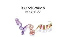
DNA Structure & ReplicationSlide2
DNA StructureSlide3
The discovery of the structure
Francis Crick and James Watson
The deoxyribonucleic acid (DNA) molecule is the genetic blueprint for each cell and ultimately the blueprint that determines every characteristic of a living organism. The DNA molecule was discovered in 1951 by Francis Crick, James Watson, and Maurice Wilkins using X-ray diffraction. Slide4
Photo Researchers, Inc.
"Francis
Crick
and James Watson," Microsoft® Encarta® 98 Encyclopedia. © 1993-1997 Microsoft Corporation. All rights reserved.Slide5
In 1953 Crick,
left,
and Watson,
right,
described the structure of the DNA molecule as a double helix, somewhat like a spiral staircase with many individual steps. In 1962 Crick, Watson, and Wilkins received the Nobel Prize for their pioneering work on the structure of the DNA molecule. Slide6
A little extra info
Although Maurice Wilkins from Cambridge is credited as a key player in the discovery, it was actually Rosalind Franklin, who was working in his lab, that used a technique called X-ray diffraction to her determine the structure of DNA – she got no credit for the discovery!Slide7
Watson/Crick Model of DNA
1. 2 chains of nucleotides coiled around each other to form a double helix.
2. The nitrogen bases of the 2 chains are joined together by weak hydrogen bonds. (easily broken)Slide8
Watson/Crick Model of DNA
3. A specific purine base is paired with a specific pyrimidine base. Adenine with Thymine (A-T) Guanine with Cytosine (G-C)
4. The sequence of base pairs along the DNA molecule determine the genetic code. Slide9
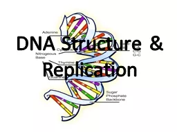
StructureSlide10
Structure
Looks like a twisted ladder
The “handrails” or backbone is made of the phosphate and 5-C (pentose) sugar called deoxyribose
The “rungs” are made of the joined nitrogen bases
The nucleotides are joined together by covalent bonds into a single strandSlide11
Nitrogen Bases
There are 4 different nitrogen bases
Adinine,Thymine,Guanine,Cytosine
As Chargaff’s rule indicates:
Adinine bonds with Thymine
Guanine bonds with CytosineTherefore there will always be equal amounts of A and T , G&C.
Slide12Slide13
Purines
Nitrogen bases with a double ring structure
Adenine and GuanineSlide14
Pyrimidines
Nitrogen bases that have a single ring structure
Thymine and Cytosine (and Uracil of RNA)Slide15
Complementary Base Pairs
A two ring base will bind with a one ring base so that there are always three rings that separate the backbone
Three hydrogen bonds attach cytosine to guanine
Two hydrogen bonds attach thymine to adenine.Slide16
Antiparallel
The two strands run opposite to each other
One end of the chain is 3’ (sugar end) the other end is 5’ (phosphate end)
Hydrogen bondsSlide17
Draw and label a simple diagram of the molecular structure of DNA
Un-seeable BiologySlide18
What is a “genome”?
the genome
is the entirety of an organism's
hereditary
information
The genome includes both the genes and the non-coding sequences of the DNA/RNA.Slide19
RNA
Not the same as DNA because:
The sugar component of RNA is ribose rather than deoxyribose
Uracil
instead of
ThymineRemains single stranded, though it can fold back on itself to produce regions of complementary base pairsSlide20
THE CENTRAL DOGMASlide21
Table 12.01Slide22
Fig. 12.08Slide23
DNA ReplicationInterphase of Mitosis/Meiosis
Semi conservative
Meaning one old strand combines with a new strand to produce two new double strands of DNASlide24
Parent Molecule
Separation of strands
“Daughter” DNA molecules each consisting of one parent strand and one new strandSlide25
Animation
http://www.mcgrawhill.ca/school/applets/abbio/ch18/dna_replication.swfSlide26Slide27
DNA Replication
semi-conservative replication
-new DNA molecule made of one parent and one newly replicated strand.
In general a DNA molecule ‘unzips’ down the middle of the paired bases, 2 individual strands are made that will become the ‘templates’ for new complete DNA standsSlide28
The Steps for DNA Replication (during S-phase of Interphase):
Initiation starts at a specific nucleotide sequence, a group of enzymes called
DNA helicases
breaks hydrogen bonds between bases to unzip the double helix
Proteins bind to keep strands apartSlide29Slide30
RNA primers
attach to a spot on the original DNA stand
DNA polymerase III
– starts at where the primer attached to the DNA and makes new strand in 5’ to 3’ direction (always)Slide31
DNA polymerase 1
– removes primers and replaces with nucleotide
DNA ligase
– joins DNA fragmentsSlide32Slide33
DNA Replication
Remember—Replication 3’-5’Slide34
Build a DNA
DNA Replication
DNA makes DNA
Un-seeable Biology
2:52