
Valvular heart disease may have congenital or acquired causes Valves on the left side are most commonly affected due to higher pressures Valvular disease is classified as Stenosis narrowed opening that impedes blood moving forward ID: 909103
Download Presentation The PPT/PDF document "Valvular heart disease Key points" is the property of its rightful owner. Permission is granted to download and print the materials on this web site for personal, non-commercial use only, and to display it on your personal computer provided you do not modify the materials and that you retain all copyright notices contained in the materials. By downloading content from our website, you accept the terms of this agreement.


Slide1
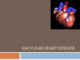
Valvular heart disease
Slide2Key points
Valvular
heart disease may have congenital or acquired causes.
Valves on the left side are most commonly affected due to higher pressures.
Valvular
disease is classified as:
Stenosis – narrowed opening that impedes blood moving forward.
Insufficiency – improper closure – some blood flows
backward (regurgitation
).
Congenital
valvular
disease may affect all four valves and cause either stenosis
or insufficiency
.
Acquired
valvular
disease is classified as one of three types:
Degenerative disease – due to damage over time from mechanical
stress; mostly results from hypertension.
Rheumatic disease – gradual fibrotic changes, calcification of valve
cusps. The
mitral valve is most commonly affected.
Infective endocarditis – infectious organisms destroy the
valve. Streptococcal
infections are a common cause.
Slide3Risk Factors for
Valvular
Heart
Disease
Hypertension
Rheumatic fever (mitral stenosis and insufficiency)
Infective endocarditis
Congenital malformations
Marfan
syndrome
Slide4Diagnostic Procedures and Nursing Interventions
Chest x-ray (chamber
enlargement, pulmonary congestion, and
valve calcification).
12-lead electrocardiogram (ECG) shows chamber hypertrophy.
Echoco
(US) show s
chamber size, hypertrophy, specific valve
dysfunction, ejection
function, and amount of
regurgitant
flow
.
Exercise tolerance
testing (stress echo);
impact
of the
valve problem on functioning during stress.
Angiography
reveals chamber pressures, ejection fraction, regurgitation,
and pressure
gradients
Slide5Therapeutic Procedures and Nursing Interventions
Percutaneous
balloon
valvuloplasty
may open the
stenotic
aortic or
mitral valves
. A catheter is inserted through the femoral artery and advanced to
the heart. A
balloon is inflated at the
stenotic
lesion to
open
the fused
commissures
and improve
leaflet
mobility
.
Surgical management includes valve repair, chordae
tendineae
reconstruction and
prosthetic valve replacement.
Prosthetic valves may be mechanical or tissue. Mechanical valves last
longer but
require anticoagulation. Tissue valves last 10 to 15 years.
Slide6Assessments
Monitor
for signs and symptoms
.
Left-sided valve damage results in
dyspnea
,
fatigue,
increased pulmonary artery pressure,
and
decreased cardiac output.
Right-sided valve damage results in
dyspnea, fatigue
, increased right atrial pressure,
peripheral edema, jugular vein distention, and hepatomegaly
Slide7Mitral stenosis
Mitral insufficiency
Aortic stenosis
Aortic insufficiency
Palpitations
Proximal nocturnal
Dyspnea
Angina
Angina
Hemoptysis
Orthopnea
Angina
S3
Hoarseness
Palpitations
Syncope
Diastolic murmur
Dysphagia
S3 and/or S4
Decreased SVR
Widened pulse
pressure
Jugular vein
distention
Crackles in lungs
S3 and/or S4
Orthopnea
Systolic murmur
Systolic murmur
Cough
Atrial fibrillation
Narrowed pulse
pressure
Diastolic murmur
Atrial fibrillation
Slide8Tricuspid stenosis
Tricuspid
insufficiency
Pulmonic stenosis
Pulmonic
insufficiency
Atrial dysrhythmias
Conduction delays
Cyanosis
Diastolic murmur
Diastolic murmur
Supraventricular
tachycardia
Systolic murmur
Decreased cardiac
output
Systolic murmur
Slide9Assess/Monitor
Oxygen
status
Vital signs
Cardiac rhythm
Hemodynamics
Heart and lung sounds
Exercise tolerance
Slide10NANDA Nursing Diagnoses
Decreased
cardiac output
Impaired gas exchange
Activity intolerance
Acute pain
Slide11Nursing Interventions
Administer O2
as prescribed to improve myocardial oxygenation.
Maintain fluid and sodium restriction.
Administer medications as prescribed.
Diuretics to decrease preload.
Antihypertensive agents (beta-blockers, calcium-channel blockers,
ACE
Inotropic agents to increase contractility – digoxin (
Lanoxin
),
dobutamine
.
Anticoagulation therapy for clients with mechanical valve
replacement
Assist
the client to conserve energy and decrease myocardial oxygen consumption.
Post-surgery care is similar to coronary artery bypass surgery (care for
sternal incision
, activity limits for 6 weeks, report fever).
Slide12Nursing Interventions
Client EducationProphylactic antibiotics are recommended prior to dental work,
surgery, or
other invasive procedures.
Encourage the client to follow the prescribed exercise
program.
Encourage adherence to dietary restrictions; consider
nutritional
consultation.
Teach the client energy conservation.
Slide13Complications and Nursing Implications
Heart
failure is the inability of the heart to maintain adequate circulation to
meet tissue
needs for oxygen and nutrients.
Ineffective
valves result in heart failure.
Monitoring a client’s heart failure class (I to IV) is often the gauge for
surgical intervention
for
valvular
problems.
Slide14Angina and Myocardial Infarction
Slide15Angina pectoris is a clinical syndrome usually characterized by episodes of pain or pressure in the anterior chest . The cause is usually insufficient coronary blood flow which results in a decreased oxygen supply to meet an increased myocardial demand for oxygen in response to physical exertion or emotional stress.
Slide16Slide17Key Points
The continuum from angina to myocardial infarction (MI) is termed
acute coronary
syndrome
. Symptoms of acute coronary syndrome are due to
an imbalance
between myocardial oxygen supply and demand.
Angina pectoris is a warning sign for acute MI.
Women and older adults may not always experience symptoms typically
associated with
angina or MI.
The majority of deaths from an MI occur within 1
hr
of symptom onset.
Early
recognition
and treatment
of acute MI is essential to prevent death.
Research shows improved outcomes following an MI in clients treated with
aspirin, beta-blockers
, and angiotensin-converting enzyme (ACE) inhibitors.
Slide18Key Points
When blood flow to the heart is compromised, ischemia causes chest pain.
Anginal
pain
is often described as a tight squeezing, heavy pressure, or constricting
feeling in
the chest. The pain may radiate to the jaw, neck, or arm.
The three types of angina are:
Stable angina (
exertional
angina) occurs with exercise or emotional
stress and
is relieved by rest or nitroglycerin.
Unstable angina (
preinfarction
angina) occurs with exercise or
emotional stress
, but it increases in occurrence, severity, and duration over
time
.
Variant angina (
Prinzmetal’s
angina) is due to coronary artery spasm,
often occurring
at rest.
Slide19Slide20Key Points
Pain unrelieved by rest or nitroglycerine and lasting for more than 15
min differentiates
MI from angina.
An abrupt interruption of oxygen to the heart muscle produces
myocardial ischemia
. Ischemia may lead to tissue necrosis (infarction) if blood supply
and oxygen
are not restored. Ischemia is reversible; infarction results in
permanent damage
.
When the cardiac muscle suffers ischemic injury, cardiac enzymes are released
into the
bloodstream, providing specific markers of MI.
Slide21Key Points
MIs are classified based on:
The affected area of the heart (anterior, anterolateral).
The depth of involvement (
transmural
versus
nontransmural
).
The EKG changes produced (Q wave, non-Q wave). Non-Q-wave MIs are
more common
in older adults, women, and clients with diabetes.
Slide22Risk Factors for Angina and MI
Male
gender
Hypertension
Smoking history
Increased age
Hyperlipidemia
Metabolic disorders: Diabetes mellitus, hyperthyroidism
Methamphetamine or cocaine use
Stress: Occupational, physical exercise, sexual
activity
Obesity
Lack of
exercise
Hx
of cardiac disease
Slide23Diagnostic Procedures and Nursing Interventions
ECG:
Check for changes on serial ECGs.
Angina: ST depression and/or T-wave inversion (ischemia)
MI: T-wave inversion (ischemia), ST-segment elevation (injury), and
an abnormal
Q wave (necrosis)
Clients with non-ST elevation MIs have other indicators.
ST segment depression that resolves with relief of chest pain
New development of left bundle branch block
T-wave inversion in all chest leads
Serial Cardiac Enzymes: Typical pattern of elevation and decrease back to
baseline occurs
with MI
.
Cardiac catheterization reveals the exact location of coronary artery
obstructions and
the degree of ischemia and necrosis
.
Slide24Slide25Slide26Therapeutic Procedures and Nursing Interventions
Percutaneous
transluminal
coronary angioplasty (PTCA) uses a balloon at
the tip
of a catheter guided under fluoroscopy to press plaque against the vessel
wall and
to
dilates
the obstructed coronary artery to increase/restore tissue perfusion.
Stents may be placed to maintain patency. Following a PTCA, monitor for
bleeding (heparin),
acute vessel closure (emergency coronary artery bypass graft),
and dysrhythmias
(reperfusion).
Coronary artery bypass graft (CABG) surgery restores myocardial tissue
perfusion by
the addition of grafts bypassing the obstructed coronary arteries.
Slide27Slide28Assessments
May
be asymptomatic
Chest pain (
substernal
/
precordial
, may radiate to the neck, arms,
shoulders or
jaw; tight squeezing or heaviness in the chest, burning, aching,
dull, constant
)
Dyspnea
Pallor and cool, clammy skin
Tachycardia and/or palpitations
Anxiety/fear, feeling of
doom
Angina is accompanied by severe apprehension and a feeling
of impending death.
Sweating (diaphoresis)
Nausea and
vomiting
A feeling of weakness or numbness in the arms, wrists, and
hands
Dizziness, decreased level of consciousness
Slide29Slide30Assessment
Angina is usually a result of atherosclerotic heart disease
and is
associated with a significant obstruction of a major
coronary artery
.
Factors
affecting
anginal
pain are physical
exertion, exposure
to cold, eating a heavy meal, or stress or any
emotion- provoking
situation that increases blood pressure,
heart rate
, and myocardial workload
.
Slide31Slide32Assess/Monitor
Vital
signs every 15 min until stable, then every hour
Serial ECG, continuous ST segment monitoring
Location, severity, quality, and duration of pain
Continuously monitor cardiac rhythm
Oxygen saturation levels
Hourly urine output – greater than 30 mL/
hr
indicates renal perfusion
Laboratory data: Cardiac enzymes, electrolytes, ABGs
Slide33NANDA Nursing Diagnoses
Ineffective cardiac tissue perfusion secondary to CAD as evidenced by chest pain or other prodromal symptoms Death anxiety
Decreased
cardiac output
Acute pain
Anxiety/fear
Activity intolerance
Deficient knowledge about underlying disease and methods for avoiding complications
Noncompliance, ineffective management of therapeutic
regimen related to failure to accept necessary lifestyle
changes
Slide34Nursing
Interventions
The objective is to decrease
O2
demand of myocardium and to increase
O2
supply
Administer
oxygen (4 to 6 L), as prescribed.
Obtain and maintain IV access.
Promote energy conservation
Administer medications as prescribed.
Vasodilators;
Nitroglycerin is
the medication
of choice.
Analgesics reduce
pain (Morphine
is the medication of
choice).
Beta-blockers
(
propranolol )
have
antidysrhythmic
and antihypertensive
Thrombolytic
agents can be effective in dissolving thrombi if
administered the
first 6
hr
following an MI.
Antiplatelet; Aspirin is the medication of choice.
Anticoagulants
Glycoprotein
IIB/IIIA inhibitors (thrombolytic agents) prevent the
binding of
fibrogen
and thus block platelet aggregation.
Slide35Teach the client to avoid straining, strenuous exercise, or emotional stress when
possible.
Client education regarding response to chest pain:
Stop activity and rest.
Place nitroglycerin tablet under tongue to dissolve (quick absorption).
Repeat every 5 min if the pain is not relieved.
Call 911 if the pain is not relieved in 15 min.
Prepare the client for diagnostic examinations as prescribed and revascularization
procedures (angiography, angioplasty, CABG).
Encourage lifestyle modifications to lower incidence of recurrence: smoking
cessation, limiting saturated fat/cholesterol, weight management, and blood
pressure control. Make appropriate referrals (for example, dietician).
Slide36Complications and Nursing Implications
Acute
MI is a complication of angina not relieved by rest or nitroglycerin.
Cardiogenic shock is a serious complication of pump failure, commonly
following an
MI of 40% or more of the left ventricle. It is Class IV heart
failure (tachycardia
, hypotension
,
inadequate urinary output (less than
30
mL/
hr
), altered level of consciousness, respiratory distress (crackles,
tachypnea),
cool, clammy skin, decreased
peripheral pulses, and chest pain.
Intervention: O2, ET, morphine IV
and/or nitroglycerin
, vasopressors IV
and/or
positive inotropes Other
possible emergency
interventions include
use of an
intra-
aortic
balloon pump
and/or emergency CABG
Ischemic mitral regurgitation due to myocardial ischemia may be evidenced
by the
development of new cardiac murmur
.
Dysrhythmias due to myocardial
hypoperfusion
require vigilant
continuous cardiac
monitoring.
Ventricular aneurysms/rupture due to myocardial necrosis may present
as sudden
chest pain, dysrhythmias, and severe hypotension.
Slide37Slide38Slide3939
Prevention
Self care action plan changing habits.
Stop smoking
Increase level of exercise
Cut down on fatty foods
Eat more oats, which decrease cholesterol
Slide40Slide4141
Lose wt if u DR. thinks you are overweight.
Make sure your BP is not high by regular check
Consider another method of contraceptive if you take pill
Slide42Slide4343
THANK YOU