
and histology Dr Gallatz Katalin PARTS AND WALLS OF THE ORAL CAVITY FLOOR OF THE ORAL CAVITY ORAL DIAPHRAGM HARD AND SOFT PALATE ISTHMUS OF FAUCES BLOOD SUPPLY OF THE ORAL CAVITY Parts ID: 917762
Download Presentation The PPT/PDF document "Oral cavity , morphology" is the property of its rightful owner. Permission is granted to download and print the materials on this web site for personal, non-commercial use only, and to display it on your personal computer provided you do not modify the materials and that you retain all copyright notices contained in the materials. By downloading content from our website, you accept the terms of this agreement.


Slide1
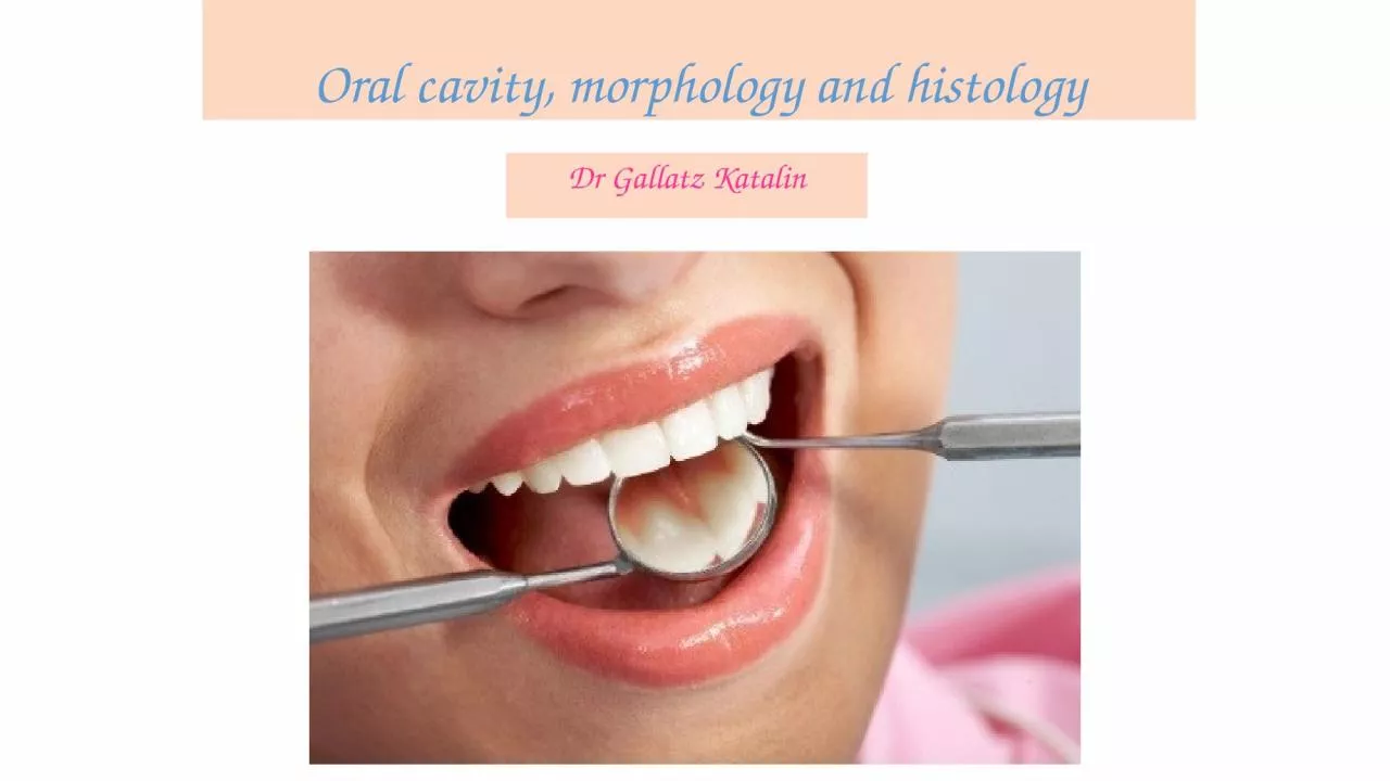
Oral cavity, morphology and histology
Dr Gallatz Katalin
Slide2PARTS AND WALLS OF THE ORAL CAVITY
FLOOR OF THE ORAL CAVITY – ORAL DIAPHRAGM
HARD AND SOFT PALATE
ISTHMUS OF FAUCES
BLOOD SUPPLY OF THE ORAL CAVITY
Slide3Parts of
the
oral
cavityVestibule –vestibulum oris outside of the dental archesOral cavity proper inside of the dental arches
Slide4Borders
of
the
vestibulum
oris :lateral: cheek (bucca) anterior: lips
medial
:
dental
arches
Borders
of
the
oral
caity
proper
:
outside
:
dental
arches
roof
:
palate
floor
:
oral
diaphragm
posterioly
:
isthmus
of
the
fauces
Slide5Borders
of
the
vestibulum
oris :lateral: cheek (bucca) anterior: lipsmedial
:
dental
arches
Borders
of
the
oral
caity
proper
:
outside
:
dental
arches
roof
:
palate
floor
:
oral
diaphragm
posterioly
:
isthmus
of
the
fauces
Slide6Floor
of
the
oral
cavity
Slide7Parts
and
floor
of
the
oral cavity
Slide8LATERAL LINGUAL SULCUS
B
orders
:
lateral
.: mylohyoid m.medial. Hyoglossus m.superior: sublingual
mucous
membrane
Content
:
-
l
ingual
nerve
-
s
ubmandibular duct- hypoglossal nerve- deep lingual vein
FLOOR OF THE ORAL CAVITY AND LATERAL LINGUAL SULCUS
Slide9BONES
PALATINE PROCESS OF THE MAXILLA
HORIZONTAL PLATE OF THE PALATINE BONE
SUTURES
MEDIAN AND TRANSVERSE PALATINE SUTURE
OPENINGS
INCISIVE CANAL
GREATER AND LESSER PALATINE FORAMINA
ROOF OF THE ORAL CAVITY - PALATE
Slide10HARD AND SOFT PALATE
palatine
raphe
:
longitudinal
mucosal
fold
in
the
midline
palatine
rugae
:several (3-7) transverse folds
(
choping
and
grinding
of
foods
,
phonation
,
sucking
)
palatinae foveolae:openings of the ducts of the small salivaryglands
Slide11BLOOD SUPPLY
-
d
escendending
palatine a. ascending palatine a. nasopalatine a.
INNERVATION:
nasopalatine
n.
greater
palatine
n.
lesser
palatine nerves
Slide12STRUCTURES OPENING INTO THE ORAL CAVITY:DUCTS OF THE BIG SALIVARY GLANDS: DUCT OF THE PAROTID GLAND
DUCT OF THE
SUBMANDIBULAR GLAND
DUCT OF THE
GLANDULA SUBLINGUAL GLANDDUCTS OF THE SMALL SALIVARY GLANDS: DUCTS OF LINGUAL GLANDS DUCTS OF LABIAL GLANDS DUCTS OF BUCCAL GLANDS DUCTS OF PALATINE GLANDS
Slide13PAROTID DUCT
-
Runs
paralel
with
the zygomatic arch at the anterior edge of the masseter turn medially and pierces the
buccinator
,
-
Opens
into
the
vestibulum
oris through the the parotid papilla at the level of the apex of
the
root
of
2.upper
molar
teeth
.
Slide14SUBLINGUAL REGION
frenulum
linguae
,
sublingual
caruncula
sublingual
fold,
fimbriate
fold,
lingual
vein
Slide15SUBMANDIBULAR DUCT – WHARTON’S DUCT
-
It
runs
in the lateral sulcus of the tongue first between the lingual and hypoglossal nerve, - anteriorly crosses
the
lingual
nerve
and
runs
above
it
, opens through the sublingual caruncula together with the major sublingual duct .
Sublingual caruncula
Slide16ISTHMUS OF THE FAUCES
Borders
:
superior
:
soft palateinferior: root of the tonquelateral: palatoglossal and
palatopharngeal
arch
POSTERIOR BORDER OF THE ORAL CAVITY
Slide17ARTERIES OF THE HEAD
EXTERNAL CAROTID ARTERY
SUP.THYROID A.
LINGUAL A
.
ASCENDING PHARYNGEAL A.FACIAL A.OCCIPITAL A.MAXILLARY A.SPF. TEMPORAL A.
Slide18BLOOD SUPPLY OF THE ORAL CAVITY
n
asopalatina
a.
sphenopalatine a.(
a.
max
.)
anterior
palate
descending
palatina
a.
maxillary
a. palateascending palatina a. facial a. palatelingual a. ext
carotid
a.
t
ongue
,
sublingual
region
buccal
a
. maxillary a cheeksup.and inf labial a. facial a. lipsinfraorbital a. maxillary a. upper
teeth
i
nf.alveolar
a.
m
axillary
a.
l
ower
teeth
Slide19MAXILLARY ARTERY
M
andibular
part:
deep
auricular
a.
ant
.
t
ympanic
a.
i
nf
. alveolaris a. middle meningeal a.Pterygoid part: masseteric a. deep temporal a. pterygoid
arteries
buccal
a.
sup
.,
post.alveolar
br . Pterygopalatine part: infraorbital a. desc.palatina a. sphenopalatine a.
Slide20BRANCHES OF THE FACIAL ARTERY
-
ascending
palatina a. submental a. glandular arteries
inferior
és
superior
labial
aa
.
angular
a.
Slide21Veins
of
the
head
and neck
Slide22Slide23Slide24Slide25ORAL MUCOSALINING MUCOSA
2. MASTICATORY MUCOSA
3. SPECIALISED MUCOSA
Slide26LINING MUCOSA covers the oral
surface of the
lips
,
cheeks, floor of mouth, and the ventral surface of the tongue - stratified squamous non-keratinized epithelium - lamina propria, - tela submucosa (small salivary
glands
:
labial
,
buccal
are
present
)
2.
MASTICATORY MUCOSA
covers the hard palate and gingiva - stratified squamous non-keratinized epithelium (at the tip of interdental papilla of the gingiva it
can
be
keratinized
)
-
lamina
propria
firmly
attaches to the
periosteum of the bones MUCOPERIOSTEUM
Slide273. SPECIALISED MUCOSAThe dorsal surface and lateral borders of
the tongue
are
covered by a specialised mucous membrane that contains different types of papillas: FILIFORM FUNGIFORM CIRCUMVALLATAE FOLIATAEFoliate and circumvallate papillas have important role in
taste
perception
1.
Tunica
mucosa
:
-
stratified
squamous
non keratinezed epithelium - lamina propria2
.
Aponeurosis
of
the
tongue
3
.
Skeletal
muscles,
glands, adipose tissueLINING MUCOSA
Slide28Foliate and circumvallate papillas have important
role in
taste
perception
, they contain gustatory receptors – taste buds
Slide29FRONTAL SECTION
MR
Slide30X-RAY
Slide31PATHOLOGY
STOMATITIS APHTOSA ACUTE TONSILLITIS
Slide32CANCER
Leukoplakia
generally refers to a firmly
attached
white patch
on a mucous membrane which is associated with an increased risk of cancer. It usually occurs within the mouth, although sometimes mucosa in other parts of the gastrointestinal tract, urinary tract, or genitals may be affected.[
Slide33Thank
you
for
your
attention
!