
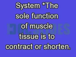
ANSC 3404 Objectives Study the structures of muscle and the mechanism of muscle contraction SKELETAL SMOOTH CARDIAC METHOD OF CONTROL VOLUNTARY INVOLUNTARY INVOLUNTARY BANDING PATTERN STRIATED NONSTRIATED STRIATED ID: 192948
Download Presentation The PPT/PDF document "Structure and Function of Muscle" is the property of its rightful owner. Permission is granted to download and print the materials on this web site for personal, non-commercial use only, and to display it on your personal computer provided you do not modify the materials and that you retain all copyright notices contained in the materials. By downloading content from our website, you accept the terms of this agreement.


Slide1
Structure and Function of Muscle
ANSC 3404
Objectives
: Study the structures of muscle and the mechanism of muscle contraction.Slide2
SKELETAL SMOOTH CARDIACMETHOD OF CONTROL VOLUNTARY INVOLUNTARY INVOLUNTARYBANDING PATTERN STRIATED NON-STRIATED STRIATEDNUCLEI/CELL MULTI SINGLE SINGLE
Muscle TypesSlide3
Cardiac MuscleSlide4
Smooth MuscleSlide5
Skeletal MuscleSlide6Slide7
Muscle Cross Sections Showing Bundles of
MyofibersFAT CELL
BLOOD VESSELSlide8
Cross Section of Muscle Fibers
NUCLEISlide9
MyofiberSlide10
Red and White Fibers in MuscleSlide11
Fiber typesSlide12
The Blood Supply for
MyofibersSlide13
Connective TissuesSlide14
Position of
Mysiums in MuscleENDO = within PERI = aroundEPI = upon
Endomysium
Perimysium
EpimysiumSlide15
Endomysium from muscle not aged Endomysium after cooler aging (28 D At 4oC)Slide16
The
Sarcoplasmic Reticulum Sarcoplasmic reticulum T-tubuleCalcium StorageRequired for contractionSlide17
Structure of MuscleSlide18
Structure of Muscle (Cont)Slide19
Sarcomere
Functional unit of a muscleRuns from z-line to z-lineActinMyosinSlide20
Myosin FilamentSlide21
Actin
FilamentSlide22
Muscle StructureSlide23
Critical Contractile ProteinsSlide24
REVIEWSlide25
Fat StructuresSlide26
ADIPOSE
TISSUEBLOOD VESSEL
ADIPOCYTE
ADIPOCYTESSlide27
Fat Layers and Depots
I.F. = Inter-fasicular or intramucular
(marbling)
I.M. =
Intermuscular
(
seam fat)
PR. =
Perinephric
or
Peri
-renal
(fat around the kidneys)Slide28
FAT CELLS
Adipoblasts develop at widely varying rates in different parts of the body.Slide29
Adipogenesis
Adipoblasts20 microns in diameterAdipocytes120 micron in diameter300 micron in obese
Cellular make-up95% of cytoplasm is lipid
Remainder primarily nucleusSlide30
ADIPOCYTESlide31
HOW ARE ADIPOCYTES FORMED?
ONCE RECRUITED & FILLED, AN ADIPOCYTE IS EASIER TO FILL AGAIN THAN IF NOT FILLED BEFOREAdipose cells begin to accumulate
When filled with lipid, it is a mature adipocyteSlide32
Muscle ContractionSlide33
Introduction
Overall structure of muscle is designed for contraction and relaxation, which leads to movement and locomotion.The ability to contract and relax is lost during the transformation of muscle to meat.Events surrounding this conversion greatly impact meat palatabilitySlide34
Introduction
The biochemical processes that provide energy to the living muscle cause the accumulation of metabolites during harvestAffects color, WHC, pH, othersAn understanding of muscle contraction is necessary to understand these processesSlide35
Contraction
Begins with stimuli that arrive at the surface of the muscle fiber at the sarcolemmaNerve impulse starts in the brain and is transmitted via nerves to the muscleSlide36
Transmembrane
PotentialsUnder resting conditions, an electric potential exists between the inside and outside of the cellFluids inside are negativeFluids outside are positiveResults in a resting membrane potentialSlide37Slide38
Transmembrane
PotentialsExtracellular – Na+ and Cl-Intracellular – K+ and A-Na+ and K+ gradient maintained by a sodium-potassium pump.Slide39
Action Potential
Transmits electric impulse to muscleTravels along the membrane surface of the nerve fiber by depolarizationInitiated by a dramatic increase in the permeability of Na+Na+ rushes into cell to establish equilibrium; however K+ stays in cell causing a change in the net charge inside the cell to positiveLasts only a millisecond (0.5 to 1 millisecond) before the permeability to Na+ is changed to resting stateSlide40Slide41
Myoneural
JunctionAction potential is not strong enough to elicit a response aloneUses a chemical transmitter called acetylcholine to be released.Acetylcholinesterase is quickly released to neutralize the acetylcholineSlide42Slide43Slide44
Muscle Action Potentials
Same as the action potential for nerve fibersCommunicated to the inner muscle cell via the T-tubule systemAction potential transverse a muscle fiber via the t-tubules and are ultimately responsible for the release of calcium from the SRSlide45
Sarcomere
- Basic contractile unit of the muscleMyosinActinTropomyosinTroponin
Z linesSlide46
Elements required for muscle contraction and relaxation
Acetylcholine and AcetylcholinesteraseCalciumAdenosine 5'-triphosphate (ATP)Derived from aerobic and anaerobic metabolismSlide47
Sources of Energy for Muscle Contraction and RelaxationSlide48
Energy
AerobicGlycolysisTCA cycleElectron transport chainAnerobicExcess Hydrogen is used to reduce pyruvic
acid to lactic acid, which permits glycolysis
to proceed at a rapid rate
Easily fatiguedSlide49Slide50Slide51
Contraction Phase
1. Nerve pulse/impulse transmitted through action potential2. Acetylcholine is released at neural juncture3. Action potential transmitted to muscle fiber via the T-tubles
to the sarcoplasmic
reticulum (SR)Slide52Slide53Slide54
Contraction phase
4. Calcium is released from SR into sarcoplasm5. Calcium binds to troponin
6. ATP is hydrolyzed (burned)
7. Energy causes a shift in
tropomyosin
and
actin
binding site is exposedSlide55Slide56
Contraction phase
8. Actin-myosin cross bridge forms (cross bridge is termed actomyosin)9. ATP hydrolyzed
10. Myosin head rotates
11. Repeated over and over; filaments slide causing shortening of
sarcomereSlide57Slide58
ContractionSlide59
A
sarcomere contractingNotice that neither filament changes length
ACTIN MYOSINSlide60
http://www.unmc.edu/Physiology/Mann/mann14.html
http://www.blackwellpublishing.com/matthews/myosin.htmlhttp://entochem.tamu.edu/musclestruccontractswf/index.htmlSlide61
Relaxation phase
1. Acetylcholinesterase is released (neutralizes acetylcholine)
2. Calcium pump activated by SR to sequester calcium
3.
Actin
-myosin cross bridge terminated
4.
Tropomyosin
shifts covering the binding site on
actinSlide62
Relaxation phase
5. Passive sliding of filaments6. Sarcomere returns to resting stateSlide63Slide64
Be Able To:
Draw a sarcomere showing: Myosin Actin Z Line A Band I Band H Zone
M Line Explain how a muscle contracts and relaxes
starting with a nerve impulse and including the SR role
Explain the role of ATP in muscle contraction and relaxation
(See p.889-900 In
The Meat We Eat
)