

1 g Teaching Kit 2 Sr No Contents 1 Aim 3 2 Introduction 3 3 Principle 3 4 Kit Contents 5 Materials Required But Not Provided 6 Storage 6 7 Important Instructions 8 Procedure 6 9 Fl ID: 940594
Download Pdf The PPT/PDF document "Day2 Inoculation of host strain from Da..." is the property of its rightful owner. Permission is granted to download and print the materials on this web site for personal, non-commercial use only, and to display it on your personal computer provided you do not modify the materials and that you retain all copyright notices contained in the materials. By downloading content from our website, you accept the terms of this agreement.


1 Day2- Inoculation of host strain from Day 3- Preparation of competent cells g Teaching Kit 2 Sr. No. Contents 1 Aim 3 2 Introduction 3 3 Principle 3 4 Kit Contents 5 Materials Required But Not Provided 6 Storage 6 7 Important Instructions 8 Procedure 6 9 Flowchart 8 10 Observation and Result 11 Interpretation 12 Troubleshooting Guide 3 To learn the process of cloning a foreign gene into a vector. The cloning experiments of Herbert Boyer, Stanley Cohen, Paul Berg, and their colleagues in the early 1970s ushered in the era of recombinant DNA technology. Recombinant DNA is a DNA that has been created artificially. DNA from two or more sources is incorporated into a single recombinant molecule. The basic procedure of molecular cloning involves a series of steps. First, the DNA fragments to be cloned are generated by using restriction endonucleases. Second, the fragments produced by digestion with restriction enzymes are ligated to other DNA molecules that serve as vectors. Vectors can replicate autonomously (independent of host genome replication) in host cells and facilitate the manipulation of the newly created recombinant DNA molecule. Third, the recombinant DNA molecule is transferred to a host cell. Within this cell, the recombinant DNA molecule replicates, producing dozens of identical copies known as clones. As the host cells replicate, the recombinant DNA is passed on to all progeny cells, creating a population of identical cells, all carrying the cloned sequence. Finally, the cloned DNA segments can be recovered from the host cell, purified, and analyzed in various ways. Principle: Cloning refers to the exact copy of an organ, whole organism, single cell or a piece of DNA. Gene cloning is a process through which an exact copy of a particular gene is made. The process through which a foreign piece of DNA is transferred into another DNA led to the development of recombinant DNA technologies. The entire cloning procedure involves the following steps: Isolation of pure vector and insert DNA Restriction digestion of the DNAs Ligation of the two linear DNA fragments Transformation of the ligated product Screening for the right clone Isolation of pure vector and insert DNA: During cloning the foreign DNA is isolated after following the specific DNA isolation procedure. The insert DNA which contains the gene of interest is obtained after restriction enzyme digestion. Several naturally occurring plasmids have been engineered to make different types of cloning vectors. Cloning vec
tors are circular DNA molecules in which DNA fragments/insert are maintained and amplified. Vectors should have the following features that make them compatible for a variety of uses in recombinant DNA procedures: Vectors are relatively small molecules; most are only 2.5 - 3 kb in size. A vector should contain an origin of replication (ori) in order to maintain its autonomous state. Marker: To facilitate selection of cells that contain the vector, a vector should carry at least one genetic marker for which an easy assay exists (e.g., ampicillin resistance, Ampr). Cloning sites: A vector should contain several different restriction endonuclease sites that allow the insertion of a gene of interest during cloning. Many cloning vectors like pUC19 contain a synthetic DNA sequence called a polylinker, which consists of 21 closely-packed RE recognition sites in a small region of DNA. 2. Restriction digestion of the DNAs:During cloning procedure an insert is added to a vector and for this reason both of them have to be cleaved by restriction endonuceases (REs). Restriction endonucleases recognize a specific, rather short, nucleotide sequence on a double-stranded DNA molecule, called a restriction site, and cleave the DNA at this recognition site or elsewhere, depending on the type of enzyme. The target site or sequence which the restriction enzyme recognizes is generally from 4 to 6 base pairs and arranged in a palindromic sequence. Generally both the vector and the foreign DNA are digested with the same RE. The restriction digested vector DNA has to be dephosphorylated to prevent self-ligation.Fig 1: Cloning of a foreign DNA fragment into a vector involves several steps 3. Ligation of two linear DNA fragments:Two linear DNA molecule ends (vector and insert) can be joined together through a process called ligation. This process involves the formation of a covalent bond between two DNA fragments (having blunt or overhanging, complementary, 'sticky' ends) by the help of an enzyme named as ligase. DNA ligase forms a phosphodiester bond between the 3 hydroxyl of one nucleotide and the 5 phosphate of another. Ligation can be directional or non-directional based upon the restriction enzyme used. When both the vector and the insert are digested with a single RE then the ligation can occur in either direction and when they are digested with two REs then ligation takes place only in one direction. 4. Transformation of the ligated product: The ligation reaction mixture is introduced into bacterial cells in a process ca
lled transformation. In this method cells are incubated in a concentrated calcium salt solution to make their membranes leaky. The permeable competent cells are then mixed with DNA (ligation Foreign DNA which contains the insert Plasmid Both the vector and the foreign DNA are digested with the same restriction enzyme Foreign DNA has two restriction sites Plasmid has one restriction Cohesive generated Cleaved DNA is ligated with the plasmid vector mix) to allow entry of the DNA into the bacterial cell. Alternatively, a process called electroporation can be used that drives DNA into cells by a strong electric current. Successfully transformed bacteria will carry either recombinant or nonrecombinant plasmid DNA. Multiplication of the plasmid DNA occurs within each transformed cell. A single bacterial cell placed on a solid surface (agar plate) containing nutrients can multiply to form a visible colony made of millions of identical cells. As the host cell divides, the plasmid vectors are passed on to progeny, where they continue to replicate. 5. Screening of the right clone:The last step during cloning is screening for the right clone. Screening means to detect the right clone among a population of colonies and the screening procedure depends upon the vector used for cloning. The E. coli plasmid pUC19 carries sites for different restriction enzymes (Multiple Cloning Site, MCS) within the -terminal coding sequence for -galactosidase of the operon. When pUC19 is transformed into a competent host cell (which has a deletion at the amino terminal end of the LacZ gene, which codes for -galactosidase), the truncated products from both complement each other and as a result enzymatically active -galactosidase is produced. This is called -complementation. The tranformants turn blue on X-gal and IPTG containing plates due to the production of -galactosidase. X-gal is a chromogenic substrate of -galactosidase and IPTG acts as the inducer for the expression of this enzyme. If an insert is cloned within the MCS of pUC19 then -galactosidase is not produced and as a result the transformants turn white on X-gal and IPTG containing plates. As a result one can directly screen for the insert containing vector (recombinant). Kit Contents: The kit can be used to clone GFP (Green Fluorescent Protein) gene in pUC19 vector and to screen for the right clone under UV light. Table 1: Enlists the materials provided in this kit with their quantity and recommended storage Sr. No. Product Materials provided Quantity Storage 5 exp
ts 1 MB104 Ampicillin 0.03 g-20 2 M1245 Luria Bertani (LB) Broth8.5 gR T 3 M1151 Luria Bertani Agar 25 gR T 4 TKC027 E. coli Host1 No.2-8 5 TKC195 Vector 0.012 ml-20 6 TKC196 Insert 0.024 ml-20 7 TKC197 Positive Control Plasmid0.012 ml-20 8 TKC198 10X Ligase Assay Buffer0.012 ml-20 9 TKC199 T4 DNA Ligase0.006 ml-20 10 TKC065 1M Calcium chloride (Sterile)20 mlR T 11 PW1139 Collection Tubes, Polypropylene (2.0 ml)18 Nos.R T 12 CG282 Polypropylene Tube (0.2 ml)7 Nos.R T Materials Required But Not Provided: Conical flask, Measuring cylinder, Beaker Other requirements: Micropipettes, Tips, 50 ml Centrifuge Tubes, Water bath (42C Incubator, 37Shaker, Centrifuge, UV Transilluminator, Crushed ice, Sterile double distilled water, Sterile loop and spreader Storage: Plasmid DNA Cloning Teaching Kit is stable for 6 months from the date of receipt without showing any reduction in performance. On receipt, store E.coli Host at 2-8C. Vector, insert, control Plasmid, 10X ligase buffer, T4 DNA Ligase and Ampicillin should be stored at 20C. All other kit contents can be stored at room temperature (15-25Important Instructions: Day 1: Open the vial containing culture and resuspend the cells with 0.25 ml of LB broth.Pick up a loopful of culture and streak onto LB agar plate with ampicillinIncubate overnight at 37 Inoculate a single colony from the revived plate in 1 ml LB broth. Incubate at 37C overnight. Thaw 10X Ligase Buffer, T4 DNA Ligase, Vector and insert on ice and then set up the ligation reaction as follows: Water : 11 Vector DNA : 2 Insert DNA Read the entire procedure carefully before starting the experiment. The entire procedure should be carried out under sterile conditions. Preparation of 0.1M Calcium chloride (Sterile) (200 ml): To prepare 200 ml of 0.1M Calcium chloride, take 20 ml of 1M Calcium chloride and add 180 ml of sterile distilled water. Store at 2-8Prior to the preparation of competent cells, pre-chm chloridesolution and centrifuge tubes. Set the centrifuge at 4C and water bath at 42Transformation should be carried out as soon as possible after the competent cells are prepared. Storage of competent cells leads to poor or no transformants. Preparation of LB (Luria Bertani) broth (55 ml): Dissolve 1.38 g of LB media in 55 ml of distilled water. Sterilize by autoclaving. Preparation of LB (Luria Bertani) agar plates (20 ml): Dissolve 0.8 g of LB agar in 20 ml of st
erile distilled water. Sterilize by autoclaving and pour on sterile petriplate. Preparation of Ampicillin: Dissolve 30 mg of ampicillin powder in 600 l of sterile double distilled water to give a concentration of 50 mg/ml. Preparation of LB (Luria Bertani) Agar plates containing Ampicillin (100 ml): Dissolve 2.5 g of LB media and 1.5 g of agar in 100 ml of distilled water. Sterilize by autoclaving and allow the media to cool down to 40-45C. Add 100 l of ampicillin, to 100 ml of autoclaved LB agar media, mix well and pour on sterile petriplates. 10X Ligase buffer : 2 T4 DNA ligase : 1 Mix the contents by tapping gently and incubate at 16C water bath, overnight. Take 50 ml of LB broth in a sterile flask. Transfer 1 ml overnight grown culture into this flask. Incubate at 37C shaker at 300 rpm for 3-4 hours till the O.D reaches ~ 0.6. Preparation of Competent Cells: Prepare competent cells within 3 days of reviving the strain. Transfer the above culture into a prechilled 50 ml polypropylene tube (not provided). Allow the culture to cool down to 4C by storing on ice for 10 minutes. Centrifuge at 4100 rpm for 10 minutes at 4C. Decant the medium completely. No traces of medium should be left. Resuspend the cell pellet in 30 ml prechilled sterile 0.1 M Calcium chloride solution. Incubate on ice for 30 minutes. Centrifuge at 4100 rpm for 10 minutes at 4C. Decant the calcium chloride solution completely. No traces of solution should be left. Resuspend the pellet in 2 ml prechilled sterile 0.1M Calcium chloride solution. 10.This cell suspension contains competent cells and can be used for transformation. Transformation of cells: l of the above cell suspension in three 2.0 ml collection tubes and label them as control, plasmid and ligation mix. Add 2 l of positive control plasmid DNA to the tube labeled as plasmid and mix well. Add 10 l of the ligation mix to the tube labeled as ligation mix and mix well. Incubate all the 3 tubes on ice for 30 minutes. Transfer them to a preheated water bath set at a temperature of 42C for 2 minutes (heat shock). Rapidly transfer the tubes on ice. Allow the cells to chill for 5 minutes. Add 800 l of LB Broth to all the tubes. Incubate the tubes for 1 hour at 37C to allow the bacteria to recover and to express the antibiotic resistance marker encoded by the plasmid. l of cell culture from each tube on LB agar plate containing ampicillin using a sterile spreader. Store at room temperature till the plates are dry. Incubate the plates overnight at 37
8 0bservation and Result: After incubation observe the plates under UV light (on a UV transilluminator) and look for the glowing green colonies on the plasmid plate. For the ligation mix plate look for both glowing and non-glowing colonies. Interpretation: On transformation of cells with positive control plasmid, antibiotic resistance is conferred on the host as this plasmid carries gene for ampicillin resistance. As a result, those cells that grow in presence of ampicillin are transformed cells. In contrast, no growth is observed on plating competent cells untransformed with vector DNA as the host E.coli cell is sensitive to ampicillin. The transformed colonies of the positive control plate glow when exposed to UV light as it contains GFP gene. The unique 3 dimensional structure of the GFP releases energy in the form of visible green light on exposure to UV light. For the ligation mix plate, the colonies which carry the recombinant plasmid (vector+insert) glow under UV light and those which carry only the vector (self-ligation) do not fluoresce under UV light. No. Problem Possible Cause Solution Transformants from the ligation mix do not fluoresce under UV light Ligation is not done properly Set up the ligation assay as mentioned in the protocol. Always thaw the ligase buffer and the enzyme on ice. Try to maintain the temperature during ligation Few or no transformants Cold chain not maintained properly during the procedure Always keep the cells on ice & make sure the centrifuge machine is cold throughout the experiment Heat shock treatment not done accurately Follow the procedure, with exact time duration for heat shock treatment used is not correct While making the plates add accurate amount of Plates were not incubated at proper temperature for sufficient time Incubate the plates at 37C for minimum 18 hours Observed on control plate Ampicillin is inactivated Add ampicillin to the media after it cools down to 40-45 Contamination plates Sterility not maintained during the experiment Make sure that the entire procedure is performed aseptically Technical Assistance: At HiMedia we pride ourselves on the quality and availability of our technical support. For any kind of technical assistance, mail at PIHTBM022_O/0117 HTBM022-01 carry only the vector (self-ligation) do not fluoresce under UV light. No. ProblemPossible CauseSolution Transformants from the ligation mix do not fluoresce under UV light Ligation is not done properly Set up the ligation assay
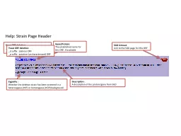
as mentioned in the protocol. Always thaw the ligase buffer and the enzyme on ice. Try to maintain the temperature during ligation Few or no transformants Cold chain not maintained properly during the procedure Always keep the cells on ice & make sure the centrifuge machine is cold throughout the experiment Heat shock treatment not done accurately Follow the procedure, with exact time duration for heat shock treatment used is not correct While making the plates add accurate amount of Plates were not incubated at proper temperature for sufficient time Incubate the plates at 37C for minimum 18 hours Observed on control plate Ampicillin is inactivated Add ampicillin to the media after it cools down to 40-45 Contamination plates Sterility not maintained during the experiment Make sure that the entire procedure is performed aseptically Technical Assistance: At HiMedia we pride ourselves on the quality and availability of our technical support. For any kind of technical assistance, mail at PIHTBM022_O/0117 8Flowchart: 0bservation and Result: After incubation observe the plates under UV light (on a UV transilluminator) and look for the glowing green colonies on the plasmid plate. For the ligation mix plate look for both glowing and non-glowing colonies. Interpretation: ion of cells with positive control plasmid, antibiotic resistance is conferred on the host as this plasmid carries gene for ampicillin resistance. As a result, those cells that grow in presence of ampicillin are transformed cells. In contrast, no growth is observed on plating competent cells untransformed with vector DNA as the host E.coli cell is sensitive to ampicillin. The transformed colonies of the positive control plate glow when exposed to UV light as it contains GFP gene. The unique 3 dimensional structure of the GFP releases energy in the form of visible green light on exposure to UV light. For the ligation mix plate, the colonies which carry the recombinant plas+insert) glow under UV light and those which 10X Ligase buffer : 2 T4 DNA ligase : 1 Mix the contents by tapping gently and incubate at 16C water bath, overnight. Take 50 ml of LB broth in a sterile flask. Transfer 1 ml overnight grown culture into this flaIncubate at 37C shaker at 300 rpm for 3-4 hours till the O.D reacheseparation of Competent Cell competent cells within 3 days of reviving the stthe above culture into a prechilled 50 ml polypropylene tube (not Allow the culture to cool down to 4C by storing on ice for 10 minuCentrifuge at 4100 rpm f
or 10 minutes at 4 the medium completely. No traces of medium should beResuspend the cell pellet in 30 ml prechilled sterile 0.1 M Calcium chloride solutiIncubate on ice for 30 minutesifuge at 4100 rpm for 10 minutes at 4 the calcium chloride solution completely. No traces of solution should Resuspend the pellet in 2 ml prechilled sterile 0.1M Calcium chloride soluti10.This cell suspension contains competent cells and can be used for transforansformation of cethe above cell suspension in three 2.0 ml collection tubes and label them as controd and ligation mix. Add 2 l of positive control plasmid DNA to the tube labeled as plasmidand mix well. Add 10 l of the ligation mix to the tube labeled as ligation mix and mix Incubate all the 3 tubes on ice for 30 minuTransfer them to a preheated water bath set at a temperature of 42C for 2 minutes (heat shRapidly transfer the tubes on ice. Allow the cells to chAdd 800 l of LB Broth to all the tubes. Incubate the tubes for 1 hour at 37nd to express the antibiotic resistance marker encoded by the plasbe on LB agar plate containing ampicillin using a sterilespreadStore at room temperature till the Incubate the plates overnight at 37 Plasmid DNA Cloning Teaching Kit is stable for 6 months from the date of receipt without showing any reduction in performance. On receipt, store E.coli Host at 2-8C. Vector, insert, control Plasmid, 10X ligase buffer, T4 DNA Ligase and Ampicillin should be stored at –20C. All other kit contents can be stored at room temperature (15-25Important Instructions: Procedure: Day 1: n the vial containing culture and resuspend the cells with 0.25 ml of LB broth.Pick up a loopful of culture and streak onto LB agar plate with ampicillinIncubate overnight at 37 ulate a single colony from the revived plate in 1 ml LB broth.C overnight.Thaw 10X Ligase Buffer, T4 DNA Ligase, Vector and insert on ice and then set up the liWater : 11 Vector DNA : 2 Insert DNA Read the entire procedure carefully before starting the experiment. The entire procedure should under sterile conditionseparation of 0.1M Calcium chloride (Sterile) (200 ml): To prepare 200 ml of 0.1M Calciumchloride, take 20 ml of 1M Calcium chloride and add 180 ml of sterile distilled water. Store at 2-8he preparation of competent cells, pre-chm chloridesoluubes. Set the centrifuge at 4C and water bath at 42ormation should be carried out as soon as possible after the competent cells are of competent cells leads to poor or no transformants.Preparation of LB (Luria Bertani) broth (55
ml): Dissolve 1.38 g of LB media in 55 by autoclavieparation of LB (Luria Bertani) agar plates (20 ml): Dissolve 0.8 g of LB agar in 20 ml ofsterile distilled water. Sterilize by autoclaving and pour on sterile on of Ampicillin: Dissolve 30 mg of ampicillin powder in 600 l of sterile double e a concentration of 50 mg/ml.Preparation of LB (Luria Bertani) Agar plates containing Ampicillin (100 ml): Dissolve 2.g of agar in 100 ml of distilled water. Sterilize by autoclaving and allow the l down to 40-45C. Add 100 l of ampicillin, to 100 ml of autour on sterile petriplates. Plasmid DNA Cloning Teaching Kit is stable for 6 months from the date of receipt without showing any reduction in performance. On receipt, store E.coli Host at 2-8C. Vector, insert, control Plasmid, 10X ligase buffer, T4 DNA Ligase and Ampicillin should be stored at –20C. All other kit contents can be stored at room temperature (15-25Important Instructions: Procedure: Day 1: n the vial containing culture and resuspend the cells with 0.25 ml of LB broth. Pick up a loopful of culture and streak onto LB agar plate Incubate overnight at 37 a single colony from the revived plate in 1 ml LB broth.C overnight.Thaw 10X Ligase Buffer, T4 DNA Ligase, Vector and insert on ice and then set up the liWater : 11 Vector DNA : 2 Insert DNA Read the entire procedure carefully before starting the experiment. The entire procedure should beunder sterile conditionseparation of 0.1M Calcium chloride (Sterile) (200 ml): To prepare 200 ml of 0.1M Calciumchloride, take 20 ml of 1M Calcium chloride and add 180 ml of sterile distilled water. Store at 2-8he preparation of competent cells, pre-chm chloridesoluubes. Set the centrifuge at 4C and water bath at 42ormation should be carried out as soon as possible after the competent cells are of competent cells leads to poor or no transformants.Preparation of LB (Luria Bertani) broth (55 ml): Dissolve 1.38 g of LB media in 55 by autoclaviPreparation of LB (Luria Bertani) agar plates (20 ml): Dissolve 0.8 g of LB agar in 20 ml ofsterile distilled water. Sterilize by autoclaving and pour on sterile on of Ampicillin: Dissolve 30 mg of ampicillin powder in 600 l of sterile double e a concentration of 50 mg/ml.Preparation of LB (Luria Bertani) Agar plates containing Ampicillin (100 ml): Dissolve 2.media and 1.5 g of agar in 100 ml of distilled water. Sterilize by autoclaving and allow the media tol down to 40-45C. Add 100 l of ampicillin, to 100 ml of autoclaved LB agar media, mix well andur on sterile petri