
fig 1 An OCTScan and fluorescein angiography fig 2 was performed showing subretinal hyperreflective material and early foveal patchy hyperfluorescence indicative of CNV We assumed CNV secondary to degenerative myopia and IV ranibizumab was administered in the OS with VA improving to ID: 930676
Download Presentation The PPT/PDF document "CASE REPORT A 42 years old woman present..." is the property of its rightful owner. Permission is granted to download and print the materials on this web site for personal, non-commercial use only, and to display it on your personal computer provided you do not modify the materials and that you retain all copyright notices contained in the materials. By downloading content from our website, you accept the terms of this agreement.


Slide1
CASE REPORT
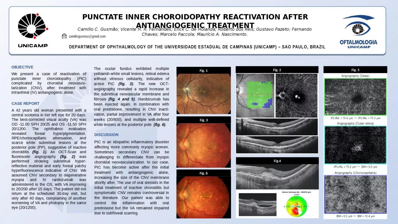
A 42 years old woman presented with a central scotoma in her left eye for 20 days. The best-corrected visual acuity (VA) was OD -11.00 SPH 20/25 and OS -11.50 SPH 20/1200. The ophthalmic evaluation revealed foveal hyperpigmentation, RPE/choriocapillaris attenuation, and scarce white subretinal lesions at the posterior pole (PP), suggestive of inactive choroiditis (fig. 1). An OCT-Scan and fluorescein angiography (fig. 2) was performed showing subretinal hyper-reflective material and early foveal patchy hyperfluorescence indicative of CNV. We assumed CNV secondary to degenerative myopia and IV ranibizumab was administered in the OS, with VA improving to 20/200 after 15 days. The patient did not return at the scheduled 30-day visit, but only after 40 days, complaining of another worsening of VA and photopsy in the same eye (20/1200).
PUNCTATE INNER CHOROIDOPATHY REACTIVATION AFTER ANTIANGIOGENIC TREATMENT
Camillo C. Gusmão; Vicente H. R. Fernandes; Erick C. de Holanda; Roberto dos Reis; Gustavo
Pazeto; Fernando Chaves; Marcelo Paccola; Maurício A. Nascimento.
DEPARTMENT OF OPHTHALMOLOGY OF THE UNIVERSIDADE ESTADUAL DE CAMPINAS (UNICAMP) – SAO PAULO, BRAZIL
camillogusmao@gmail.com
OBJECTIVE
We present a case of reactivation of punctate inner choroidopathy (PIC) complicated by choroidal neovascu-larization (CNV), after treatment with intravitreal (IV) antiangiogenic alone.
The ocular fundus exhibited multiple yellowish-white small lesions, retinal edema without vitreous cellularity, indicative of active PIC
(fig. 3)
. The new OCT-angiography revealed a rapid increase in the subretinal neovascular membrane and fibrosis
(fig. 4 and 5). Ranibizumab has been injected again, in combination with oral prednisone, resulting in CNV inacti-vation, partial improvement in VA after four weeks (20/800), and multiple well-defined white lesions at the posterior pole (fig. 6).
DISCUSSION
PIC is an idiopathic inflammatory disorder affecting more commonly myopic women. Sometimes secondary CNV can be challenging to differentiate from myopic choroidal neovascularization. In our case, PIC has become active after the initial treatment with antiangiogenic alone, increasing the size of the CNV membrane shortly after. The use of oral steroids in the initial treatment of inactive choroiditis but symptomatic CNV remains controversial in the literature. Our patient was able to control the inflammation with oral prednisone but the VA remained impaired due to subfoveal scarring.
Fig. 1
Fig.4
Fig. 5
Fig. 3
Fig. 2
Fig. 6