/
Unit  II Lecture  2:  Plants Unit  II Lecture  2:  Plants

Unit II Lecture 2: Plants - PowerPoint Presentation

topslugger
topslugger . @topslugger
Follow
342 views
Uploaded On 2022-08-02

Unit II Lecture 2: Plants - PPT Presentation

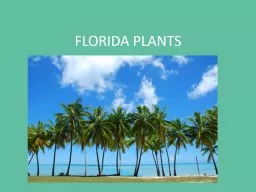
producing Photosensitization Dr Kumari Anjana Asstt Prof cum Jr Scientist Deptt Of Vety Pharmacology and Toxicology BVC BASUPatna VPT609 20 Toxicology of Plants and Toxins ID: 932735

due photosensitization lantana sheep photosensitization due sheep lantana skin animals photodynamic liver plant plants toxin ingestion lantadene bile secondary

Share:

Link:

Embed:

Download Presentation from below link

Download Presentation The PPT/PDF document "Unit II Lecture 2: Plants" is the property of its rightful owner. Permission is granted to download and print the materials on this web site for personal, non-commercial use only, and to display it on your personal computer provided you do not modify the materials and that you retain all copyright notices contained in the materials. By downloading content from our website, you accept the terms of this agreement.


Presentation Transcript

Slide1

Unit IILecture 2: Plants producing Photosensitization

Dr. Kumari AnjanaAsstt. Prof. cum Jr. ScientistDeptt. Of Vety. Pharmacology and ToxicologyB.V.C, BASU,Patna

VPT-609 (

2+0

)

Toxicology of Plants and Toxins

Slide2

Plants producing photosensitization

Slide3

PhotosensitizationPhotosensitization: is a syndrome of abnormal sensitivity of the superficial layers of unpigmented or light skinned areas of the body (nose, face, back, udder, testes, teats, mucosa,

corneae etc.) to ultraviolet (UV) and visible light probably due to the presence of some abnormal substances, termed as photodynamic agent, in the peripheral circulation.The reaction is limited to body areas which receive direct sunlight (teats and udder in cows; head, face, ears, mandibular area and cornea in sheep and goats etc.

Slide4

Severity of the condition depends on -the amount of photodynamic substance consumed, duration of ingestion and time of exposure to sunlight.This syndrome may be observed after a few hours to few days of ingestion on exposure to strong sunlight.Thus, photosensitization generally involves a triad of hepato- toxic plant, fresh – green feed in the diet and exposure to bright sunlight.

Slide5

All most all species of animals, namely bovines, ovines, caprines, equines and birds are affected . Canines and felines are rarely affected.Depending upon the circumstances how photodynamic substances accumulates in the body- by ingestion of the preformed photosensitizing agents, abnormal metabolism and/or reduced excretion of phylloerythrin (a product of chlorophyll breakdown), due to hepatic damage and

metabolic defects in porphyrin metabolism.

Slide6

Photosensitization has been categorized into three types namely: Primary photosensitization, Secondary photosensitization and Inherited or congenital photosensitization. The first two types are commonly encountered in animals.

Slide7

Primary PhotosensitizationIt is due to ingestion of exogenous agents like, drugs/ chemicals or plants containing photodynamic agent. Such agents are inherently photodynamic/photosensitive (get activated by UV radiation of 400 nm). They are mainly of plant origin from livestock point of view.

On consumption of these plants, active principles is released in the stomach, comes to the peripheral circulation in the capillaries under skin and on exposure to sunlight, theses sensitize skin similar that by porphyrins.

Slide8

Some plants causing primary photosensitization (photodynamic principle) are:Parthenium hysterophorum (parthenin), Fagopyrum

esculentum (Beech wheat) (Fagopyrin), Hypericum perforatum (st. John’s Wort) (Hypericin), Chemicals (Dyes): Acridine and rose bengal.Drugs: Phenothiazine

(Sheep: Phenothiazone

sulfoxide: Photosensitization keratitis).

Slide9

Secondary PhotosensitizationIt is a consequence of liver disease (hepatitis) swollen hepatocytesmicroobstruction of intrahepatic biliary

ductules obstruction of common/hepatic bile duct blockade of secretion of bile into intestines.

Slide10

It is therefore secondary to liver pathology. It is of plant origin. Normally chlorophyll, the photosynthetic pigment of plants, ingested though green greases by the livestock is excreted in faeces. However, due to obstruction of bile ducts/flow (above) phylloerythrin is not excreted and it enters in peripheral circulation causes photosensitization.

Slide11

Some examples of plants causing hepatotoxicity and secondary photosensitiztion are:Agave spp Lantana camara Shobia

arabecaLupinus augustifolium Senecio jacobaeum Tribulus terrestris etc.In addition all the drugs or chemicals (CCI4) causing liver damage are expected to cause secondary photosensitization in grazing or greens fed livestock.

Slide12

Congenital photosensitizationIt is often hereditary in origin due to certain enzymatic abnormalities or inadequate production of enzymes like catalase, glucose-6-phosphate dehydrogenase etc. in RBCSs and disruption of haeme

biosynthetic pathway. As a result, porphyrin or its derivatives accumulated in the body including skin. These substances in skin when get exposed to bright sunlight get excited, interact with cellular macromolecules or molecular oxygen to generate toxic free radicals.

Slide13

Congenital porphyria is a metabolic defect due to abnormality in haeme synthesis: ferroprotoporphyrin has been observed in cattle, swine and cats. As a result, protoporphyrin, uroporphyrin II and coproporphyrin

III accumulate in higher concentrations in serum and tissues. Accumulation in dentine and bones give rise to red colour and results in pink tooth or osteohaemochondrsis. There is increased excretion of porphyrins in urine (porphyrinuria) which is reddish brown in colour. Kidneys and lungs of affected animals also fluoresce in UV light. Sometimes anaemia due to inadequate biosynthesis of haemoglobin and reduced life span of erythrocytes is also observed.

Slide14

Genesis of photosentitization reaction or syndromeIn all the three types the genesis of photodynamic reaction or photodynamic syndrome is almost similar, though the causes are different as explained above. The mechanism is explained as follows: The photosensitive or photodynamic agent while come in contact with ultra violet radiation of a specific wave length (< 400 nm)

and combines with molecular oxygen to form oxygen radicals.

Slide15

These oxygen free radicals injure or lyse the lysosomal membrane releasing the destructive lysosomal enzymes into the cytoplasm of the cells and eventually causing necrosis of the tissues underlying the skin. Histamine is released in the damaged tissues causing local reaction in the skin such as intense itching/

pruritis, erythema, oedema, oozing serous fluid and pain. The affected skin areas become highly sensitive to secondary bacterial infections. If not treated gangrene sets in with sloughing of the affected skin areas.

Slide16

Clinical signsThe dermal signs start with erythema, itching, oedema (swelling) and irritation. Serous fluid oozes from the area due to rubbing against fixed objects. Necrosis, gangrene and exfoliation or sloughing of the superficial skin layers. Conjunctivitis and lacrimation (watery to thick) in cattle. In sheep blindness may result due to corneal clouding/opacity. In

hepatogenous type there is jaundice. The swollen mandibular area (oedematous) in sheep looks like bottle jaw and the head looks big (big head syndrome).

Slide17

Break of wool and high mortality is also characteristic in sheep.Compared to sheep, conjunctivitis is more pronounced while oedema is less marked in cattle.In congenital type increased excretion of porphyrins in urine (porphyrinuria: reddish-brown urine).The symptoms in equines are similar to that of cattle and sheep except colic, inflammation of the mucus membrane of mouth and brain.

Slide18

Lantana camera IntroductionLantana camara

(common name: lantana, wild sage, bunch berry) is very hardy shrub and is one of the ten most toxic weeds in the world.It was brought to India in early part of the 19th century as an ornamental plant. Now, this plant has found throughout the country, hills and plains. There are several varieties (distinguished by the colour of flowers), the red variety is considered most toxic. It can propagate rapidly by means of stumps or cuttings.Natural propagation

: from seeds disseminated by the birds through their droppings or the faeces of moving flocks of sheep and goats.

Slide19

Grazing livestock normally do not eat the plant due to very pungent odour.Ingestion of leaves is the cause of toxicity. Animal affected: All ruminant : cattle, buffalo, sheep, goat.Non ruminant: horses, rabbit guinea pig, female rat. Lantana camera affect animal husbandry in two ways ;

Morbidity: loss of fodder allelopathic effect “double edges weapon” Mortality: due to ingestion of lantana foliageAllelopathic action: It causes growth inhibition of the neighbouring vegetation, due to which is rapidly changing the ecological balance. contd.

Slide20

Toxic principlesLantana hepatotoxins are – pentacyclic triterpenoids called lantadenes. Some major lantadenes are: lantadene A, lantadene B, lantadene C, lantadene D. All lantadenes have a common core structure of 22- hydroxyl-oleanonic

acid. Minor lantadenes : reduced lantadene A and reduced lantadene B are also present in lantana leaves. The toxicity is characterized by hepatotoxicity, cholestasis and photosensitization.

Slide21

Mechanism of toxicosisIngestion of lantana foliage - hepatotoxicity and secondary photosensitization. Lantana manifest its toxicity in animals in three phases viz. Gastrointestinal (GIT) phase, Hepatic phase and Post hepatic phase.

Gastrointestinal (GIT) phase:Ingestion of lantana plant.Absorption toxin from GIT (small intestine). Metabolism of lantadene A and BHepatic phase and Post hepatic phase:Toxins are transported to liver by portal blood. Toxin interact with hepatocytes and causes intrahepatic cholestasis.It causes paralysis of gall bladder and closure of bile canaliculi. Cholistasis causes regurgitation of bile which cause increase in level of bilirubin and phylloerythrine.

Slide22

Ingestion of plant by the animals Digestion of the plant in the rumen Absorption of the toxin from the rumen and small intestine Transportation of the toxin to the liver in the portal blood Metabolism of the toxin in the liver Secretion of the metabolites into the bile Metabolites injure the bile canalicular membranes Cholestasis

contd.

Slide23

Retention of bilirubin Retention of phylloerythrin Ruminal stasis Retention of toxin (rumen) Absorption of the toxin from the rumen

Cholestasis

Photosensitization

Jaundice

=

Further injury to the liver

contd.

Slide24

Post mortem lesions:The liver is ochre coloured and greatly swollen.The gall bladder is greatly distended.The rumen contents are usually dry and undigested.There is impaction of faeces in the colon.Adrenals are enlarged and the thickened cortex turns yellow.

Diagnosis: History.Clinical signs.Clinical pathology.Post mortem lesions.

Slide25

TreatmentStop further exposure of the animals to noxious weed.Keep the animals in well shaded areas.Administer IV excessive amounts of glucose saline solution.Give hepatoprotective agents to tone up the liver.Remove the toxic ruminal contents by rumenotomy

.Replace the ruminal contents with a suspension containing electrolytes, chaffed forage and rumen liquor from a healthy animals.Antibiotic and antihistaminic therapy.Administer a single dose of activated charcoal (5g/kg) to bind the toxin in the rumen and prevent further absorption.Aversion therapy involves conditioning of animal before they are left for grazing in pasture infested with poisonous plants.Vaccination: Abs against lantadene A & B could be detected in sheep and cattle after injection of the conjugates of these compounds with suitable proteins. In vaccinated animals – severity of toxicity less.