
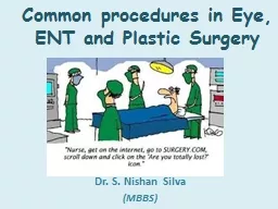
Dr S Nishan Silva MBBS The basic eye exam Snellens Chart Ophthalmoscope Slit lamp Case 1 Chalazion Treatment warm compresses lid hygiene surgical incision and curettage ID: 910502
Download Presentation The PPT/PDF document "Common procedures in Eye, ENT and Plasti..." is the property of its rightful owner. Permission is granted to download and print the materials on this web site for personal, non-commercial use only, and to display it on your personal computer provided you do not modify the materials and that you retain all copyright notices contained in the materials. By downloading content from our website, you accept the terms of this agreement.


Slide1
Common procedures in Eye, ENT and Plastic Surgery
Dr. S. Nishan Silva(MBBS)
Slide2The basic eye exam
*
*
Snellen’s
Chart ; Ophthalmoscope ; Slit lamp
Slide3Case 1
Slide4Chalazion
Treatmentwarm compresseslid hygienesurgical incision and curettagesteroid injectionpathological examination for suspicious lesion
Slide5Chalazion
Slide6Pterygium
Slide7Cataract
Slide8Slide9Cataract surgery is typically an outpatient procedure that takes less than an hour
Most people are awake and need only local anesthesia On rare occasions some people may need general anesthesia if they have difficulty laying flat or have claustrophobia
Slide10Slide11Slide12Two things happen during cataract surgery — the clouded lens is removed, and a clear artificial lens is implanted
Slide13PhacoemulsificationDuring phacoemulsification, phaco for short, the surgeon makes a small incision, where the cornea meets the conjunctiva
Slide14The surgeon then uses the probe, which vibrates with ultrasound waves, to break up (emulsify) the cataract and suction out the fragments
Slide15Once the cataract is removed, a clear artificial lens is implanted to replace the original clouded lens
This lens implant is made of plastic, acrylic or silicone and becomes a permanent part of the eye
Slide16Some IOLs are rigid plastic and implanted through an incision that requires several stitches (sutures) to close
However, many IOLs are flexible, allowing a smaller incision that requires no stitches
Slide17Phaco+IOL surgeryFoldable IOL insertion
Slide18Typical injectable IOL
Superflex lens: 6.25mm x 12.50mm
C-Flex lens 5.75mm x 12.00 mm
Slide19Cflex/Superflex injector
Slide20Loading the Superflex IOL
Slide21Insertion of AC-IOL
If adequate capsular support absent
2. Peripheral
iridectomy
3. Glide insertion
4. Coating of IOL
with viscoelastic
substance
5. Insertion of IOL
6. Suturing of
incision
1. Constriction of pupil
Slide22Patients usually go home the same dayPatients are seen in the office the next day, the following week, and then again after a month so that he or she can check the healing progress
It's normal to feel mild discomfort for a couple of days after surgeryYou may wear an eye patch or protective shield the day of surgeryYour doctor may prescribe medications to prevent infection and control eye pressure
Slide23Post-op CoursePatients are usually examined 1 day, 1 week and then one month after the surgery date
Slide24Complications of SurgeryVitreous Loss- 3.1%Vitreous Hemorrhage-0.3%Uveitis-1.8%Increased Eye Pressure- 1.2%
Retinal Detachment- 0.7%Endophthalmitis- 0.13%
Slide25Post Operative PeriodContact your doctor immediately if you experience any of the following signs or symptoms after cataract surgery:
Vision loss Pain that persists despite the use of over-the-counter pain medications A definite increase in eye redness Light flashes or multiple spots (floaters) in front of the eye
Nausea, vomiting or excessive coughing
Slide26Diabetic Retinopathy
Diabetic retinopathy is the most common cause of new cases of blindness among adults 20-74 years of age.
Each year, between 12,000 to 24,000 people lose their sight because of diabetes.
During the first two decades of disease, nearly all patients with type 1 diabetes and over 60% of patients with type 2 diabetes have retinopathy
Slide27A classification of diabetic retinopathy
A useful classification according to the types of lesions detected on fundoscopy is as follows:
Non-proliferative diabetic retinopathy (NPDR)
Mild non-proliferative diabetic retinopathy
Microaneurysms
Dot and blot haemorrhages
Hard ( intra-retinal ) exudates
Moderate-to-severe non-proliferative diabetic retinopathy The above lesions, usually with exacerbation, plus:Cotton-wool spots
Venous beading and loopsIntraretinal microvascular abnormalities ( IRMA )
Proliferative diabetic retinopathy Neovascularization of the retina, optic disc or iris
Fibrous tissue adherent to vitreous face of retina
Retinal detachment
Vitreous haemorrhage
Pre retinal haemorrhage
Maculopathy
Clinically significant macular oedema (CSME )
Ischaemic
Maculopathy
Slide28Pathogenesis of Diabetic Microangiopathy
Hyperglycaemia causes-BM thickening
non
enzymaitc
glycosylation
increased free radical activity
increased flux through the
polyol pathwayosmotic damage
Haemostatic abnormalities of the microcirculation-.
Slide29Non-proliferative diabetic retinopathy (NPDR)
Slide30Cotton Wool Spots
Slide31Hard exudates ( Intra-retinal lipid exudates )
Accumulations of lipids leak from surrounding capillaries and microaneuryisms, they may form a circinate pattern.
Slide32Ischaemic Maculopathy
Slide33Proliferative diabetic retinopathy
Slide34Proliferative diabetic retinopathy
Slide35Proliferative diabetic retinopathy
Slide36Hypertension
Slide37Panretinal laser photocoagulation
Slide38Iris Neovascularisation
Slide39Panretinal laser photocoagulation for proliferative DR
Slide40Diabetic retinopathy
Slide41Laser Eye Surgery
Slide42What is LASIK?
LASIK stands for Laser-Assisted In
Si
tu
K
eratomileusis and is a procedure that permanently changes the shape of the cornea, the clear covering of the front of the eye, using an excimer laser.
LASIK is the most advance form of laser vision correction that is currently available.
Slide43Problems Corrected By Surgery
Myopia
Hyperopia
Astigmatism
Slide44LASIKLaser-Assisted
In Situ KeratomileusisUses a knife, called a microkeratome, to cut a flap in the cornea and an eximer laser to reshape the exposed stroma (the middle layer of the cornea)
Keratomileusis, a predecessor
to LASIK, involved removing
a section of the cornea,
reshaping it, and then
replacing it
Slide45ProcedureContact lenses change the shape of the cornea for up to several weeks after they’re worn. Glasses, therefore, must be worn for 2-4 weeks before the initial visit as well as in the time before surgery.
The contours of each eye are mapped out in a computerized topographical analysis, to provide a detailed plan for what will be removed during surgery. The thickness of the cornea will also be measured.
Slide46Before surgery, analgesic drops will be administered to numb the eye. The area around the eye will be cleaned, and a speculum will be inserted to keep the eye open.A ring will be applied to create suction to the cornea. The microkeratome is then attached to the ring and a flap is cut into the cornea.
The ring and microkeratome are removed and the flap is folded back to expose the sclera. The laser is then positioned over the eye, whereupon the patient must fixate upon a red light.Pulses of energy will destroy the pre-selected areas of tissue. Any debris are then washed from the eye, and the flap is returned to its original position.A clear shield is placed over the eye, and the patient is then allowed to leave, returning for check-ups 1
day, 1 week, 2 weeks, 3 months,
6 months, and 1 year after
surgery
Slide47How LASIK is Performed
Step 1. A suction ring is centered over the cornea of the eye
Slide48Step 2: The microkeratome creates a partial flap in the cornea of uniform thickness
Slide49Step 3: The corneal flap is folded back on the hinge exposing the middle portion of the cornea.
Slide50Step 4: The excimer laser is then used to remove tissue and reshape the center of the cornea.
Slide51Step 5: In the final step, the hinged flap is folded back into its original position.
Slide52Slide53Slide54AfterwardsFor the first few months after
surgery, visual acuity will fluctuateVision will stabilize in 3-6 months, but during that period of stabilization glares, halos, and difficulty driving at night may persistIf touch-ups are needed, they should be done in 3 months time, when the vision is fairly stabilized and a new flap need not be cut. Instead, the old one can simply be pried up. If done later, the progress of the healing will require a new flap to be cut
Slide55Corneal Transplantation
Slide56Financial DisclosureI have no financial interest in the subject matter presented
Slide57Corneal Opacity
Corneal scarring from firework accident
Slide58Corneal Clouding
Granular stromal dystrophy
Fungal keratitis
Slide59Corneal Clouding
Slide60Corneal DonationsNational Eye Bank of Sri Lankhttp://www.nationaleyebank.lk
/Tel : Hotline 2915 and 0112267266Eye donors society Sri Lankahttp://www.eyedonation.slt.lk/
Slide61Storage MediaOptisol GS allows for storage up to 10 days. Allows surgery to be scheduled electively
D to P (death to preservation) preferably less than 12 hours
Slide62Surgery: Full Thickness Surgery
Recipient tissue removed
Donor tissue sutured into recipient
Smooth Surface with only
endothelial disease
Full thickness block
of tissue removed just
to get to the endothelium
Central trephine cut
made
Sutures create an
irregular surface
with astigmatism
and blurring
Slide63Penetrating keratoplasty
Slide64Common Ear Conditions
Slide65Ear Drum-normal
Slide66Ear Wax
Wax is produced in the outer half of the ear canal and migrates outwards along with the canal skin. Inappropriate instrumentation can cause impaction.Wax impaction can cause hearing loss, pain, tinnitus, vertigo, or chronic cough but not usually discharge.
Sudden expansion after getting water in can cause sudden deafness or pain, but needs careful exclusion of other pathology behind it e.g. cholesteotoma
Be mindful of other possibilities
FB(crayon) in a child’s ear
Slide67Otitis ExternaInfection of the external auditory canal. Mediterranean ear/Swimmers ear
Usually unilateralGradual onset pruritis, pain, hearing loss, and ear discharge which varies in consistency and colour. Discharge not mucoid in consistency as no mucin glands are present in the ext aud canal. The pt is usually well. Can result in a featureless ext aud canalRisk factors: trauma, water, Immunosuppression, eczema
Can be fungal- spores might not always be visible
If treatment fails or otitis externa recurs
frequently consider sending an ear swab
for bacterial and fungal microscopy
and culture
Slide68Syringing / Irrigation
Slide69Otitis Media
Can be acute or chronicCan be with or without serous effusion (acute or chronic)Can be Acute or chronic suppurativeCan co-exist with Otitis externa
Otitis media with serous effusion= Glue Ear
Slide70Acute Otitis Media
Common in childrenUnwell/pyrexia, otalgia/dischargethere may be tenderness over the mastoiddischarge in meatusloss of outline of drum and landmarksTM: red, bulging,oedematous or perforation. Mostly viral but can be Streptococcus/Haemophilus
Risk factors:
Passive smoker
Male
Family history of otitis media. In day care
On formula feed
Slide71AOM (pus behind the eardrum)
Slide72Serous Otitis Media
Slide73Otitis media+effusion-Glue earFeatures
Dull retracted TMMay show air-fluid levelConductive hearing loss(whisper test, Rinne/weber tests)NotesCommon in children; often after AOM and can persist for weeksReduced hearing noticed by parents/teacherUnsteadiness- child falling over
80% clear at 8 weeks
Slide74Chronic Otitis MediaRecurrent ear discharge
Hearing loss, painlessPerforation of the TM – centralPresence of cholesteatomaMarginal, Attic perforationOffensive discharge, bleeding, granulations
Complications:
.
Vestibular symptoms
. Facial palsy
. Intracranial complications
Slide75Myringotomy
Slide76Slide77Cholesteotoma
Slide78Cholesteatoma
Cholesteatoma is "a three dimensional epidermoid structure exhibiting independent growth, replacing middle ear mucosa, resorbing underlying bone, and tending to recur after removal." There is usually a persistent or recurrent scanty cream coloured offensive discharge and progressive hearing loss due to ossicular
destruction or toxin induced sensory hearing loss
.
Slide79EpistaxisManagementPain meds, lower BP, calm patientPrepare ! (gown, mask, suction, speculum, meds and packing ready)
Evacuate clotsTopical vasoconstrictor and anestheticIdentify source
Slide80EpistaxisManagement
Anterior Sites- Pressure +/- cautery and/or tamponade - all packs require antibiotic prophylaxis
Slide81EpistaxisPosterior Packing
Need analgesia and sedationrequire admission and 02 saturation monitoring
Slide82Plastic Surgery
Slide83Introduction
Plastic surgery is defined as any procedure used to correct or restore either form or function to a body part.It deals with body modification and reconstructive surgery as well as surgery for aesthetically pleasing purposes.
Slide84Techniques and Procedures
Slide851) Skin Grafting
A skin graft is the replacement of a patient’s skin.Required after major skin loss from a burn, major trauma or infection (i.e. flesh eating bacteria).Usually plastic surgeons are called in to do skin grafts.
They plan their cut lines on the patients and close and remove sutures or staples in a particular sequence in order to minimize scarring.
Slide86Padgett Dermatome
Slide87Slide88Slide89Slide90Slide91Slide922)Reconstructive Surgery
It is performed to correct function, but in some cases may be used to generate a more normal appearance.Common procedures include tumour removal, facial reconstruction, hand repair, breast reduction and breast reconstruction (after a mastectomy).
Slide933) Microsurgery
The reconstruction of missing tissues usually by the transfer of tissue from another part of the body.Called microsurgery because the doctor uses a microscope in order to see the vessels and fibres he/she needs to connect after the tissue has been transferred.
Slide944) Cosmetic Surgery
Deals with enhancement of appearance for non-medical reasons.Includes any “lifting”, augmentation or implant insertion.Nose jobs, face lifts, Botox, collagen injections, breast augmentation and tummy tucks are the most common.
Brazilian Butt lifts are starting to challenge though. ;)
Slide955) Body ModificationSimilar to cosmetic surgery, it is the deliberate altering of the human body for non-medical reasons.
The difference is that it may not be done for a more pleasing appearance.
Slide965) Body Modification
Includes:Any piercings or tattoos.Genital modification including circumcision.Binding procedures like corsetry, foot-binding, etc…
Strange things like neck rings, “elfing”, bifurcation of the tongue…
Slide97Slide98