
The Manual Therapy Institute Anatomy amp Physiology Going back to Anatomy Connective Tissue Covering Endoneurium encompasses the axon or nerve fiber important role in protecting against transmission of substances across the membrane the bloodnerve barrier ID: 912054
Download Presentation The PPT/PDF document "Adverse Neural Tissue Tension" is the property of its rightful owner. Permission is granted to download and print the materials on this web site for personal, non-commercial use only, and to display it on your personal computer provided you do not modify the materials and that you retain all copyright notices contained in the materials. By downloading content from our website, you accept the terms of this agreement.


Slide1
Adverse Neural Tissue Tension
The Manual Therapy Institute
Slide2Anatomy & Physiology
Slide3Going back to Anatomy…
Slide4Connective Tissue Covering
Endoneurium
- encompasses the axon or nerve fiber; important role in protecting against transmission of substances across the membrane (the blood-nerve barrier)
Perineurium
- surrounds each fascicle; provides a
perineural
diffusion barrier capable of controlling flow of substances bi-directionally.
Epineurium
- outermost connective tissue; highly vascular and provides no diffusion barrier function
Slide5Slide6Slide7Nerve Nutrition
Bi-directional Nutritional Flow (
Axoplasmic
Flow):
Antegrade
Flow:
Fast 400mm day, carries substances used in the transmission of impulses (neurotransmitters and transmitter vesicles)
Slow 1-6 mm/day, carries substances needed for the maintenance of the structure of the axon
Retrograde Flow: 200 mm/day, responsible for carrying extracellular materials from the nerve terminal and
trophic
messages about the status of the nerve and the target tissues
Nutritional Requirements – 20% of O
2
consumption while only composing 2% of total body weight
Slide8Continuity of Central & Peripheral Nerves
The system is considered continuous in three ways:
1) the connective tissues are continuous
2) the neurons are interconnected electrically
3) the system is continuous chemically.
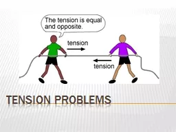
Any stresses that are imposed on the peripheral nervous system are conveyed to the central nervous system, and the reverse holds true.
Slide9Nerve Innervation
The connective tissues of the peripheral nerves, nerve roots and autonomic nervous system have a source of intrinsic
innervation
=
nervi
nervorum
Free nerve endings have been found in the:
Perineurium
Epineurium
Endoneurium
Slide10MOI’s & Examination of the Peripheral Nerve
Slide11Motor Problems
– motor neuron (body, axon, motor end plate, muscle fiber).
Signs
:
distal weakness
decreased DTR’s
myotomal
patterns
Sensory Problems
– sensory neuron (cell body in ganglion, axon, sensory receptor).
Reports
: tingling, burning,
dysesthesias
,
paresthesias
; dermatome pattern.
Mixed Nerves
– both
ANS
– sweating and/or vascular control; skin changes
Dysfunctions of Peripheral Nerves
Slide12Entrapment/Compression
- small amount of pressure chronically endured over time (
ie
posture, repeated compression or dysfunctional movement); CTS,
Cubital
Tunnel, TOS
Trauma
– laceration, severing, blunt, crushing
Heredity
– Charcot-Marie-Tooth
Nutritional/Metabolic
- diabetics, alcohol
Infections
–
Guillain-Barre
, post-Polio, Herpes Zoster, Bell’s Palsy or Trigeminal
Exposure to Toxins
– lead, organophosphates
Motor End Plate Disorders
- Myasthenia Gravis, Botulism
Both MOI’s will lead to classic nerve s/s…
Overview of MOI’s for Peripheral Nerves
Slide13Create an “at risk” environment for the neural tissue.
Be mindful of theses conditions, and whether they are under control…
Systemic Risk Factors
Slide14Microvascular diseases
Diabetes
Thyroid issues
Renal Disease
Inflammatory arthritis
Gender
Pregnancy
Obesity
Age
Smoking
Occupational exposure/activities
Systemic Risk Factors
Slide15Goals:
know the major peripheral nerves
understand the individual motor and sensory function of each peripheral nerve
be able to establish a treatment plan based upon clinical presentation…
Realize that Compression leads to:
decreased vascular flow
interrupts axonal transport and conduction
leads to myelin thinning
epineural
thickening
Nerve Compression Injuries
Slide16Diagnostic Considerations with Peripheral Nerve Compression….
Mimics some
tendonosis
/tendonitis and can occur concurrently with such
Concurrent with many other orthopedic injuries:
Lateral ankle sprains (
sural
or
peroneal
)
Proximal Humeral fractures (radial)
Knee scope (
saphenous
)
Spine
hypermobility
Occur frequently after fractures
Slide17Pt. Hx
, physical exam and laboratory data assist in diagnosis and locating lesion.
However, no one test is 100% specific or sensitive (so look at multiple pieces of the puzzle…)
Examination Considerations
Slide18Nerve Compression Diagnostic Procedures
Pt. History
Motor Exam –
myotomal
as well as specific to suspected peripheral nerve
Sensory Exam –
dermatomal
as well as specific peripheral nerve
NTPT (neural
tissue
provocation testing)
Provocative Testing (
ie
Tinel’s
,
Phalen’s
,
Roo’s
)
Physical findings - atrophy, clawing, etc.Body diagramsEMG/NCV studies
Slide19Slide20Dermatomal Key Points
Slide21Sensory Regions
Slide22Always consider proximal points of compression….
Be mindful that initial changes may be transient, but if situation persists or worsen, the changes can become permanent with fibrosis.
Localization and correct diagnosis allow for appropriate intervention planning.
Based on peripheral nerve anatomical organization, which is affected first - motor or sensory?
Examination Note:
Slide23Sidenote
on EMG/NCV
Sometimes the only objective measure
Can localize lesion by “inching”
Not always positive in early stages
Operator dependent
Assists in diagnosis and allows measurement of progression/resolution
Slide24Adverse Neural Tissue Tension
Never-ending Acronyms:
ANTT =
Adverse Neural Tissue Tension
ULTT =
Upper Limb Tension Testing
NTPT =
Neural Tissue Provocation Testing
Slide25Important Concepts
Initially
may not suspect
ANTT with straight forward orthopedic conditions
tend to
develop gradually
as a secondary result from injury
The nerve as a
source of pain
Concept of
AIG
= abnormal impulse generating site
Slide26AIG’s
Coined by David Butler
When a peripheral nerve is injured, it can develop the ability to repeatedly
& spontaneously
generate its own impulse
Slide27Main Characteristics of AIG’s
Mechano-sensitivity:
stimulated by mechanical stimuli (movement, touch, etc)
Spontaneous Activity
Susceptible Sites =
area of myelin damage or regenerating axon sprouts
Slide28When to suspect
ANTT?
When not responding as should within the expected time frame
Describes in terms consistent with ANTT - “burning”, “crawling”, “electrical”, “ants on me”, “pulling”, bizarre sounding things like “warm water”
Worsening despite objective improvement of ROM, strength, etc
Slide29Symptoms of ANTT
Development of pain &
paresthesia
is
gradual
(neural zone)
Symptoms
radiate
(either proximally or distally)
Pain along the
nerve pathway
or spot pain (
hyperalgesic
response to palpation)
Aggravated by
positions or movements
that “stretch” the nerve
Nocturnal s/s not uncommon
Slide30Signs of ANTT
Sensory nerve?
Motor nerve?
Positive neural tissue provocation testing (NTPT)
TTP along the nerve
Slide31What makes a NTPT positive?
Reproduction of
s/s (know it is relevant)
Response is altered by a
distant
component (either a distal or proximal component)
Difference in response from side to side, or what is
normal
May have to differentiate of a positive test is relevant or not….
Slide32Susceptible Sites
sites of nerve branching
unyielding interfaces
sites of nerve attachment
soft tissue and fibro-osseous tunnels
sites at which a nerve is
cutaneous
Slide33Common MOI’s
External forces –
ie
casts, belts, walking boots, ill-fitting shoes
Internal forces –
ie
swelling, bone spur
Chronic repeated
microtrauma
–
ie
posture
Double crush –
ie
ask about old injuries proximal & distal to site
Slide34Double Crush
Proteins and cell bodies travel distally while waste products travel proximally thru axonal transport systems.
Disruption causes decreased threshold for s/s or AIG’s elsewhere along the nerve.
Either site may be asymptomatic without the second insult.
What does this tell you must be done on evaluation?
Slide35ANTT Differential Diagnosis
Lumbar Radiculopathies:
Pain with cough, sneeze, Valsalva?
Well delineated area of sensory change?
Partial weakness, decreased reflexes?
Electrodiagnostic testing?
What is the
key
to differential diagnosis of ANTT and lumbar root?
Slide36The Key…
Identify a
different peripheral nerve
with the major contribution from the
same root level
as the suspected nerve
Or test a more
proximal branch
originating from the same peripheral nerve
Then compare motor and sensory function
Slide37Common LE Entrapments
Slide38Femoral
Nerve
MOI:
pelvic fracture, scarring after abdominal surgery, tumors, inguinal hernias
S/S:
most pronounced at the knee, knee buckling may occur
Local tenderness in the groin, pain and
paresthesiae
over the
anteromedial
surface of the thigh and the medial surface of the leg.
Decreased sensation over
anteromedial
thigh, weakness of quadriceps (compensated for by hyper extending the leg in standing and walking) and
sartorius
, decreased patellar tendon reflex.
Increased pain at
endrange
hip flexion and hip extension. Positive neural tension signs.
Radiculopathy
? What level? Will present with anterior tib
and adductor weakness.
R/O:
hip arthritis and
psoas
strain
Slide39Femoral &
Saphenous
Nerve
Slide40Lateral Femoral
Cutaneous
Nerve
Originates from L2 and L3, runs through the pelvis and angles downward at the ASIS.
MOI
: weight distribution changes, such as a pendulous abdomen, scarring following surgery and to direct external pressure (corset or belt)
Sites of entrapment
:
fascia
lata
inguinal ligament (naturally kinked as it passes through the ligament)
C/O
: pain, numbness and
paresthesiae
over anterior and lateral surfaces of the thigh.
Aggravating Factors
: worsened by erect posture, prolonged standing, hip extension and adduction.
There are no motor signs.
Differential diagnosis
(Differentiate from L2, L3 nerve root radiculopathy and femoral nerve neuropathy): peripheral entrapment may cause complete anesthesia with well delineated borders, while with lumbar nerve root problems the loss of sensation is usually partial due to overlap of the dermatomes)Pain over the inguinal ligament is not common with lumbar
radiculopathy
, but happens frequently with peripheral nerve entrapment of the lateral femoral
cutaneous
nerve.
Slide41Obturator
Nerve
Originates
: L2 and particularly
L3, 4
; formed in the
psoas
, runs through the pelvis to the
obturator
canal, leaves the pelvis and enters the medial thigh.
Site of entrapment =
obturator
canal.
MOIs
: pelvic fracture, hip surgery and inflammation of the pubic bone following surgery.
Clinical findings:
Pain in the groin, radiating from the inner thigh to the knee.
Decreased sensation over the medial thigh and knee, and local pain with pressure over the inguinal ligament.
Pain on
endrange
hip flexion, hip extension and abduction. Weakness in the adductor musculature leads to gait pattern disturbances: circumduction during swing phase. Duchenne during stance phase.Differential diagnosis (Radiculopathy from L2-4 and femoral nerve neuropathy) L2, 3 dermatomes include portions of the medial thigh, which could confuse the diagnosis of peripheral entrapment, but generally cover more lateral portion of the thigh. L4 is distinctively different from the sensory distribution of the
obturator
nerve.
Direct pressure in the groin should not cause pain in a lumbar
radiculopathy
, but highly typical with
obturator
entrapment.
Rule out hip arthritis and
psoas
strain.
Slide42Obturator
Nerve
Slide43Saphenous
MOI:
knee arthroscopy, medial
meniscal
repair, trauma
Slide44Saphenous
Medial Knee
Slide45Sciatic
MOI:
sacral dysfunction with
piriformis
spasm, thick wallets, scarring from hip surgery, injections,
gluteal
or pelvic tumors
Key:
glutei help differential from
radiculopathy
Caution:
Neurodynamic
testing will give the same result as for
radiculopathy
Slide46Sciatic Nerve and Major Branches
Slide47LE Nerves Commonly Involved
Tibial
:
MOI:
plantar
fascitis
,
eversion
sprains, “joggers” foot
Slide48Tarsal Tunnel Syndrome
Entrapment of posterior tibial nerve within canal:
Tibial Nerve divides into:
Medial and lateral plantar nerves
Medial calcaneal branch
Symptoms:
Pain/parathesia plantar aspect of foot
Causes:
Not clear concensus regarding causitive factors but suspect:
Excessive Pronation
Assessory FDL
Tight flexor retinaculum
Ganglion cysts
Soft tissue tumors
Slide49Tarsal Tunnel Syndrome
Slide50Anterior Tarsal Tunnel Syndrome
Deep Peroneal Nerve entrapment:
Beneath inferior extensor retinaculum
Over talonavicular joint
Over tarsometatarsal joints
Signs and Symptoms:
Pain/parathesia over dorsum to 1
st
web space
Weakness of EDB
Slide51Deep Peroneal Nerve
Slide52Plantar Nerves
Slide53LE Nerves Commonly Involved
Peroneal:
MOI:
ankle sprains, casts, walking boots, shoe wear
S/S:
pain & paresthesia lateral leg & dorsum of foot; deep peroneal - loss of sensation first web space; loss of strength in the lateral & anterior compartments
Differential:
anterior compartment syndrome (extreme pain, pallor, loss of dorsalis pedis pulse, foot drop)
Slide54Slide55Slide56Lateral Nerves at the Foot
Slide57Medial Nerves of the Foot
Slide58Adverse Neural Tissue Tensioning
Provocative Testing
Positive:
Different from uninvolved LE
Reproduction of s/s
Changes with “remote” component
Caution!
Assessment of
irritability
is key, as well as severity.
Irritability
guides the nerve glide
prescription
related to intensity…
Slide59Principles of Management of ANTT
Intensity directly related to the level of irritability present – greater the irritability the less intense the glide (
ie
NOT into s/s)
Neurological s/s (
ie
tingling, numbness) should NOT persist after gliding technique or should be improved
Choose one component motion of the testing position to utilize as the “gliding force”
Either “floss” or “glide” – either one works
Ask regarding s/s after each treatment and document changes across the course of treatment
Any worsening at all need to be immediately reported to the PT – pt. education…
Slide60Guidelines for Prescription
Educate pt on condition and goals of treatment
Emphasize NOT to push treatment – MUST be gentle
ALWAYS start with assisting to ensure perfect form and determine response; when pt masters, give nerve glides as HEP.
1. Intensity & Duration:
Initially: Perform 20-30 glides just out of s/s
Progress to: glides with minimum s/s at the end of the “glide” only
2. Frequency:
Initially: 1x day, determine response
Progress to: Gradually build up to 3x-5x day if appropriate response achieved.