

Consultant and assistant professor Pathology Objectives Know the risk factors for the development of breast cancer Know the classification of breast cancer Understand the behavior and spread of breast cancer ID: 1047597
Download Presentation The PPT/PDF document "Breast Cancer Dr Mohammed Alswayyed" is the property of its rightful owner. Permission is granted to download and print the materials on this web site for personal, non-commercial use only, and to display it on your personal computer provided you do not modify the materials and that you retain all copyright notices contained in the materials. By downloading content from our website, you accept the terms of this agreement.


1. Breast CancerDr Mohammed AlswayyedConsultant and assistant professorPathology
2. ObjectivesKnow the risk factors for the development of breast cancer.Know the classification of breast cancer.Understand the behavior and spread of breast cancer.Know the prognostic indicators of breast carcinoma.2
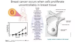
3. Introduction3Carcinoma of the breast is one of the most common cancer in women.Women who live to age 90 years, have a one in eight chance of developing breast cancerMammographic screening has dramatically increased the detection of small invasive cancers. DCIS by itself is almost exclusively detected by mammography, so the incidence of DCIS is increased with the use of mammography. Therefore number of women with invasive/advanced cancer is markedly decreased.The mortality rate have started to decline. Currently only 20% of women with breast cancer are expected to die of the disease.
4. Risk FactorsThe etiology of breast cancer in most women is unknown but most likely is due to a combination of genetic, hormonal and environmental risk factors. The major risk factors are hormones and genetics i.e. family history. Breast carcinomas can, therefore, be divided into sporadic cases, possibly related to hormonal exposure, and hereditary cases, associated with a family history or germ-line mutations.4
5. Risk FactorsHereditary breast cancerA family history of breast cancer in a first-degree relative.About 25% of familial cancers (or around 3% of all breast cancers) can be attributed to two autosomal-dominant genes: BRCA1 and BRCA2.Sporadic breast cancerThe major risk factors for sporadic breast cancer are related to exposure to hormones, gender, age at menarche and menopause, reproductive history, breast-feeding, and exogenous estrogens.The majority of these cancers occur in postmenopausal women and in cases of overexpression to estrogen.5
6. Risk FactorsAge: It increases the incidence in women. Breast cancer is rare before 25 years, except in familial forms, but the majority of cases occur in women >50 years of age. Estrogen exposure: Factors associated with exposure to increased levels of estrogen have been shown to increase a woman’s risk for breast cancer. These factors include:Early age at menarche: the younger the age at menarche, the higher her risk of breast cancer.Late age at menopause.Nulliparity.Late age at first child-birth: the earlier a woman has her first birth, the lower her lifetime risk for breast cancer. A woman who has her first birth after 30 years has an increased risk. Also postmenopausal hormone replacement slightly increases the risk.6
7. Risk FactorsFirst-degree relative with breast cancer:Women with history of cancer in first degree relative (mother, sister, aunt or daughter) are at higher risk of breast cancer. The risk increases with the number of affected first degree relatives. At least two genes that predispose to breast cancer have been identified—BRCA 1 and 2Majority of cancers occur in women without such history. Race and geographic influence: incidence of breast cancer is lower in African American women. Generally Caucasians have the highest rate of breast cancers. Breast cancer is more common in Western industrialized countries than in developing countries. 7
8. Risk FactorsRadiation exposure: Higher rate of breast cancer.History of breast cancer: Women who have had a breast cancer or have cancer in one breast are at increased risk of developing a second primary breast cancer.History of other cancers: women who have a history of ovarian or endometrial cancer are at high risk.Certain breast diseases: As noted previously women with certain types of benign breast diseases are at risk. Dietary factors e.g. high fat intake and excessive alcohol consumption.8
9. Risk FactorsObesity: may play a role.Exercise: some studies showed decreased risk with exercise.Breast feeding: the longer the women breast fed, the lower the risk.Environmental toxins: pesticides.Tobacco: Not associated with breast cancer, but associated with the development of periductal mastitis or sub-areolar abscesses.9
10. Classification of Breast CancerAlmost all (majority) are adenocarcinoma.There are two major types: Ductal LobularBreast cancer is divided into:Carcinoma in situ (non-invasive) Invasive carcinoma10
11. Carcinoma in SituThis is an epithelial proliferation that is still confined to the TDLU and has not invaded beyond the basement membrane and is therefore incapable of metastasis.There are two subtypes: Ductal carcinoma in situ (DCIS) or intraductal carcinoma (80%).Lobular carcinoma in situ (20%).11
12. Ductal Carcinoma in SituDCIS is the non-invasive proliferation of malignant cells within the duct system without breaching the underlying basement membrane.They have a very high risk of developing a subsequent invasive carcinoma.The tumor distends and distorts the ducts.Age range: same age range of invasive breast carcinoma.Often multifocal: malignant cells can spread widely through the ductal system without breaching the basement membraneWomen with DCIS are at risk of recurrent DCIS following treatment.12
13. Ductal Carcinoma in SituDCIS frequently shows micro-calcifications on mammography. Mammography is a very sensitive diagnostic procedure for detecting DCIS since majority of DCIS are not palpable. Less frequently they can present as a mammographic density or a vaguely palpable mass or nipple discharge.Because of mammography there has been a marked increase in the detection and diagnosis of DCIS in the last two decades.13
14.
15. Ductal Carcinoma in SituDifferent patterns/subtypes of DCIS can be seen e.g. comedo (central necrosis); cribiform (cells arranged around “punched-out” spaces); papillary, micropapillary and solid (cells fill spaces) DCIS can be of different grades i.e. low, intermediate and high grade 15
16. Ductal Carcinoma in SituComedo DCIS: is characterized by large central zones of necrosis with calcified debris. This type of DCIS is most frequently detected as radiologic calcifications. Less commonly, the surrounding desmoplastic response results in an ill-defined palpable mass or a mammographic density.16
17.
18. Ductal Carcinoma in SituCribriform DCIS comprises cells forming round, regular ("cookie cutter") spaces. The lumens are often filled with calcifying secretory material.18
19.
20. Ductal Carcinoma in SituSolid PatternMicropapillary Pattern20
21. Ductal Carcinoma in SituClinical behavior: may vary depending on the subtype and the gradeComedocarcinoma has essentially a 100% chance of becoming invasive if left untreated. Pure cribriform/micropapillary carries only a 30% chance of becoming invasive carcinoma. Treatment: Wide local excisionmastectomy21
22. Paget’s DiseasePaget's disease of the breast is a rare type of breast cancer that is characterized by a red, scaly eczematous lesion on the nipple and surrounding areola.Paget’s disease may be subtle or appear as an eroded and weeping erythematous eruption. Pruritus is common and it might be mistaken for eczema.Malignant cells are called Paget cells and are found scattered in the epidermis.22
23. Paget’s Disease23
24. Paget’s Disease24
25. Paget’s DiseaseThe histologic hallmark of Paget’s disease of the nipple is the infiltration of the epidermis by large neoplastic ductal cells with abundant cytoplasm, pleomorphic nuclei and prominent nucleoli. The cells usually stain positively for mucin.Paget cells extend from DCIS within the ductal system into nipple skin without crossing the basement membrane.Palpable mass can be seen in 50% of women with Paget disease indicating an underlying invasive carcinoma near by.25
26. Paget’s Disease26
27. Lobular Carcinoma in SituLCIS alone is always an incidental finding in breast biopsies performed for another reason. LCIS does not form a palpable mass and cannot be detected clinically on palpation or on gross pathological examination. Microcalcifications in LCIS are infrequent and so mammography is not useful for detection. LCIS is uncommon.LCIS tends to be multicentric and bilateral and therefore subsequent carcinomas can occur both breasts. Clinical behavior If LCIS is left untreated, about 30% of women develop an invasive cancer within 20 years of diagnosis. The invasive cancer that develops is usually lobular (but can be ductal too). LCIS is a marker of increased cancer risk in both breasts27
28. Lobular Carcinoma in SituHistology: monomorphic population of small, rounded cells fills and expands the acini of lobules. The underlying lobular architecture can still be recognized.28
29. Invasive Breast CarcinomaInvasive breast carcinoma is a tumor that has extended across the basement membrane. This permits access to lymphatics and vessels therefore the potential to metastasize.Invasive breast carcinoma is subdivided into:Ductal 80% (NOS= no otherwise specified)Lobular 10%Tubular 6%Mucinous(Colloid) 2%Medullary 2%Papillary 1%Metaplastic Carcinoma 1%29
30. Clinical Features of Invasive Breast CancerPalpable mass.About half of the patients will have axillary lymph node metastases.Larger carcinomas may be fixed to the chest wall or cause dimpling of the skin. Lymphatics may become involved and the lymphatic drainage of that area and the overlying skin gets blocked causing lymphedema and thickening of the skin, a change referred to as peau d'orange. When the tumor involves the central portion of the breast, retraction of the nipple may develop. 30
31. Clinical Features of Invasive Breast Cancer31
32. Clinical Features of Invasive Breast CancerOn mammography, invasive carcinomas commonly present as a density.Invasive carcinomas presenting as mammographic calcifications without an associated density are usually very small in size.The term "inflammatory carcinoma" refers to the clinical presentation of a carcinoma extensively involving dermal lymphatics, resulting in an enlarged erythematous breast. The diagnosis is made on clinical grounds and does not correlate with a specific histologic type of carcinoma 32
33. Clinical Features of Invasive Breast Cancer33
34. Invasive Ductal Carcinoma, NOSInvasive ductal carcinoma, NOS, is the most common type of breast cancer, forming up to 80% of these cancers.Most of these tumors induce a marked fibroblastic (desmoplastic) stromal reaction to the invading tumor cells producing a palpable mass with a hard consistency (scirrhous carcinoma). And therefore a palpable mass is the most common presentation. The tumor shows an infiltrative attachment to the surrounding structures and may cause dimpling of the skin (due to traction on suspensory ligaments) or nipple retraction. 34
35. Invasive Ductal Carcinoma, NOS35
36. Invasive Ductal Carcinoma, NOSGrossly: tumor is firm, hard, with an irregular borders.Cut surface: gritty and shows irregular margins with stellate infiltration (sometimes it can be soft and well demarcated) and in the center there are small foci of chalky white stroma and occasionally calcifications which have characteristic grating sound when cut or scraped.IDC is usually accompanied by varying amounts of DCIS.36
37. Invasive Ductal Carcinoma, NOS37
38. Invasive Ductal Carcinoma, NOSHistology: the tumor cells are large and pleomorphic usually within a dense stroma. They are adenocarcinomas and so they show glandular formation but can also be arranged in cords or sheets of cells. The tumors range from well differentiated to moderate or poorly differentiated.Carcinomas associated with a large amount of DCIS require large excisions with wide margins to reduce local recurrences38
39. Invasive Ductal Carcinoma, NOS39
40. Invasive Lobular CarcinomaIt is the second most common type of invasive breast cancer forming up to 10% of breast cancers.The tumor may occur alone or in combination with ductal carcinoma. It tends to be bilateral and multicentric.The amount of stromal reaction to the tumor varies from marked fibroblastic (desmoplastic) response to little reaction and therefore the presentation varies from a discrete mass to a subtle, diffuse indurated area. Most are firm to hard with irregular margins40
41. Invasive Lobular CarcinomaHistology: single infiltrating malignant cells, forming a line often one cell width (called as Indian file pattern). No tubules or papillary formation.41
42. Medullary CarcinomaThis subtype of breast cancer presents as a well circumscribed mass. May be mistaken clinically and radiologically for a fibroadenoma.It does not produce any fibroblastic (desmoplastic) reaction and therefore it is soft and fleshy. Histology: the tumor is composed of solid sheets of malignant cells surrounded by many lymphocytes and plasma cells. There is scant fibrous stroma. 42
43. Medullary Carcinoma43
44. Colloid Carcinoma/ Mucinous carcinoma It tends to occur in older women. It is sharply circumscribed, lacks fibrous stroma and it is slow growing.It is soft and gelatinous and has a glistening cut surface.It may be in pure mucinous or mixed with another type of invasive breast carcinoma. The tumor is composed of small islands of tumors cells and single tumor cells floating in pools of extracellular mucin44
45. Colloid Carcinoma/ Mucinous carcinoma45
46. TreatmentWide local excision (lumpectomy)MastectomySimpleModified radicalRadical46
47. Prognostic Factors - MajorInvasive or In situ disease: Invasive carcinoma has poorer prognosis as it can metastasize. In-situ carcinoma is confined to the ductal/lobular system and cannot metastasize, so it has better prognosis. Distant metastasis: Once distant metastases is present, cure is unlikely, although long-term remissions and palliation can be achieved. Favored sites for dissemination are the lungs, bones, liver, adrenals, brain, and meninges. Lymph node metastasis: Axillary lymph node status is the most important prognostic factor for invasive carcinoma. The clinical assessment of nodal involvement is very inaccurate, therefore, biopsy is necessary for accurate assessment. Tumor Size: The size of the carcinoma is the second most important prognostic factor. The risk of axillary lymph node metastases increases with the size of the carcinoma. Note: all the above parameters are used to stage the tumor. Stage is a combination of size, lymph node status and distant metastasis. Tumor size less than 2 cm is associated with a favorable prognosis. The single most important prognostic indicator is the lymph node status. Negative lymph nodes have the best prognosis. Involvement of 1 to 3 lymph nodes has an intermediate prognosis and 4 or more positive nodes have the worse prognosis.47
48. Prognostic Factors - MajorLocally advanced disease: Tumors invading into overlying skin or underlying skeletal muscle are frequently associated with concurrent or subsequent distant disease. With increased awareness of breast cancer detection, such cases have fortunately decreased in frequency and are now rare at initial presentation.Inflammatory carcinoma: Women presenting with the clinical appearance of breast swelling and skin thickening have a poor prognosis.48
49. Prognostic Factors - MinorHistologic Subtype: Infiltrating ductal and lobular carcinomas have the worst prognosis. Medullary and mucinous have intermediate prognosis. And tubular and cribriform have the most favorable prognosisTumor Grade: It is calculated using a grading system called modified Scarff-Bloom-Richardson (SBR) grading system. There are three grades: 1, 2 and 3. Grade 1 has better prognosis and grade 3 has poorer prognosis. This SBR grading system is based on the estimation of the amount of well formed glands, the degree of nuclear pleomorphism, and the mitotic rate, on microscopic examination. It is calculated by the pathologist. Tumor cells with estrogen and progesterone positive receptors: majority of breast carcinoma cells express estrogen and progesterone receptors. Such hormone positive cancers have better prognosis. They respond well to specific drugs e.g. Tamoxifen. Therefore it is mandatory to identify which tumors are ER/PR positive as they respond well to treatment and have better prognosis when compared to ER/PR negative tumors.49
50. Prognostic Factors - MinorHER2 (human epidermal growth factor receptor 2): is a glycoprotein overexpressed in about 30% of breast carcinomas. Many studies have shown that overexpression of HER2 is associated with a poor prognosis. In addition, ongoing studies have shown that HER2-overexpressing tumors respond very well to a chemotherapy drug named Trastuzumab (Herceptin). Therefore, it is mandatory to determine the HER2 status of the tumor when reporting breast cancer in order to help decide the chemotherapy plan.Lymphovascular invasion: is strongly associated with the presence of lymph node metastases and is a poor prognostic factor. Proliferative rates: ki67 index (the higher the ki67 proliferative index, the more aggressive the tumor is)50
51. ReferencesKumar V, Abbas AK, Aster JC. Robbins Basic Pathology. 10th ed. Elsevier; 2018. Philadelphia, PA.51
52. End of LectureThank You