
Marsh GA Haining J Hancock TJ Robinson R Foord A Barr JA et al Experimental Infection of Horses with Hendra VirusAustraliaHorse2008Redlands Emerg Infect Dis 2011171222322238 httpsdoiorg103201eid1712111162 ID: 1009224
Download Presentation The PPT/PDF document "Figure 4 Figure 4. Lymphadeniti..." is the property of its rightful owner. Permission is granted to download and print the materials on this web site for personal, non-commercial use only, and to display it on your personal computer provided you do not modify the materials and that you retain all copyright notices contained in the materials. By downloading content from our website, you accept the terms of this agreement.


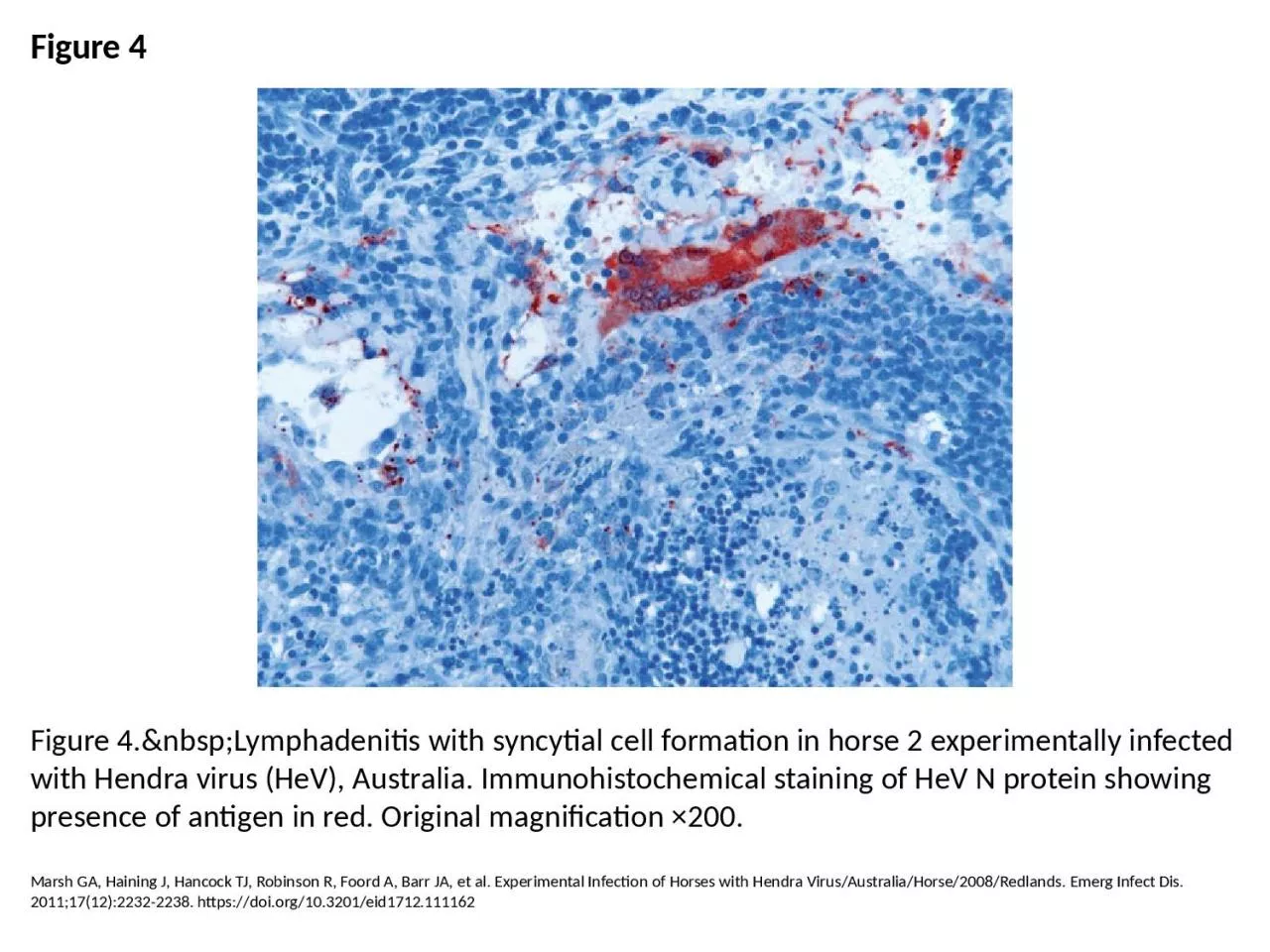
1. Figure 4Figure 4. Lymphadenitis with syncytial cell formation in horse 2 experimentally infected with Hendra virus (HeV), Australia. Immunohistochemical staining of HeV N protein showing presence of antigen in red. Original magnification ×200.Marsh GA, Haining J, Hancock TJ, Robinson R, Foord A, Barr JA, et al. Experimental Infection of Horses with Hendra Virus/Australia/Horse/2008/Redlands. Emerg Infect Dis. 2011;17(12):2232-2238. https://doi.org/10.3201/eid1712.111162