
Section 1 Overview of the Digestive System pp 852854 Two groups of organs 1 Alimentary canal also known as gastrointestinal or GI tract digests amp ID: 578002
Download Presentation The PPT/PDF document "Ch 23: The Digestive System" is the property of its rightful owner. Permission is granted to download and print the materials on this web site for personal, non-commercial use only, and to display it on your personal computer provided you do not modify the materials and that you retain all copyright notices contained in the materials. By downloading content from our website, you accept the terms of this agreement.

Slide1
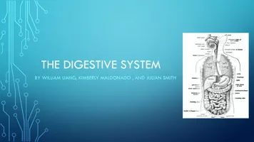
Ch 23: The Digestive System
Section 1: Overview of the Digestive System (pp. 852-854)Slide2
Two groups of organs: 1) Alimentary canal
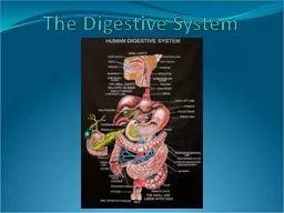
- also known as gastrointestinal or GI tract - digests & absorbs food Major organs: - mouth, pharynx, esophagus, stomach, small intestine, & large intestine
Digestive System OverviewSlide3
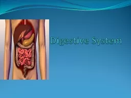
Two groups of organs: 2) Accessory digestive organs
- teeth, tongue, & gallbladder Major digestive glands: - salivary glands, liver, & pancreasDigestive System OverviewSlide4
Digestion - the physical grinding & chemical
breakdown
of food ALL digestive systems must accomplish the following:Ingestion - placing food into digestive tractMechanical breakdown - food physically broken down into smaller pieces - allows digestive enzymes to work more efficientlyDigestive ProcessesSlide5
Chemical breakdown - particles exposed to digestive enzymes - breaks
large
molecules into smaller subunitsAbsorption - subunits transported out of digestive system…into cellsElimination - indigestible material expelled from bodyDigestionSlide6
Four basic layers of tissue: 1) Mucosa
-
inner lining of canal - secretes mucus, digestive enzymes, & hormones - absorbs nutrients - protects against infectious diseasesHistology of Alimentary CanalSlide7
Four basic layers of tissue: 2) Submucosa
- dense connective tissue under mucosal layer - rich in blood supply, lymph tissue, & nerves - elastic fibers allow stomach to regain normal shape after large mealHistology of Alimentary CanalSlide8
Four basic layers of tissue: 3) Muscularis
externa
- responsible for peristalsis (rhythmic muscular contractions that move food through the canal) Composed of two layers: a) Inner circular layer opens/closes
canal b) Outer longitudinal layer lengthens
/
shortens
canal
Histology of Alimentary CanalSlide9
Four basic layers of tissue: 4) Serosa - protective,
outermost
layer - known as the visceral peritoneumHistology of Alimentary CanalSlide10
Ch 23: The Digestive System
Section 2: Functional Anatomy of the Digestive System (pp. 858-895)Slide11
Mechanical & chemical breakdown of food: - begins in the mouth
- mouth lined with thick
stratified squamous epithelium to withstand considerable friction - breakdown of food carried out mostly by the teethMouthSlide12
Function of teeth: - to break food down into smaller
fragments
- vastly improves rate & efficiency of chemical breakdown of food in stomachTeethSlide13
Tooth “Timeline”: 1) Deciduous teeth (“
baby
teeth”) - begin appearing at 6 months; complete by age 2 - 20 total - between age 6-12, roots are reabsorbed; teeth fall out 2) Permanent teeth - 32 total - All except
wisdom teeth appear by end of adolescence - Wisdom teeth appear by age
25
(sometimes never)
TeethSlide14
Teeth 1) Incisors - front of mouth; snip off pieces
of food
2) Canines - pointed; useful for tearing 3) Premolars & Molars - back of mouth; flattened surfaces used to grind food into a pasteMouthSlide15
Functions of the tongue:- reposition & mix food
during chewing- initiates swallowing response- provides ability to manipulate sounds to form words- provides sense of tasteTongueSlide16
Features of the tongue: 1) Filiform
papillae
- cone-shaped; provide friction for manipulating foods - smallest & most numerous - contain keratin; gives them a whitish appearance 2) Fungiform papillae
- scattered across surface of tongue - contain blood
vessels
; gives them a
reddish
appearance
TongueSlide17
Features of the tongue: 3) Circumvalate
papillae
- form V-shaped row on back of tongue 4) Foliate papillae - located on the sides of the tongue; near the back**Fungiform, Circumvalate, & Foliate papillae together form the taste buds**Note:
Foliate taste buds only function during infancy & early
childhood
Tongue
Super TastersSlide18
Salivary Glands - provide first stage of chemical
digestion
- pour out saliva in response smell, feel, & taste - can even be activated by the THOUGHT of foodSalivaCancerous salivary glandSlide19
Saliva - mixture of mucus & water
- contains
Amylase (digestive enzyme that begins breaking down starches into sugars) - kills bacteria & lubricates food - taste is a direct result of food dissolving in saliva
Saliva
Donuts….Slide20
After chewing… - tongue (a muscle) presses food backward into pharynx
Pharynx
- muscular cavity connecting mouth w/ esophagus & larynx - separates food/liquids from air that is being inhaledSwallowingSlide21
Epiglottis - flap of cartilage that closes off the larynx when you swallow…prevents
food
from going into lungsSwallowingSlide22
Once past the epiglottis… - Food travels into the esophagus
- Esophagus is
collapsed when emptyEsophagusSlide23
Stomach - Expandable bag…capable of holding
2-4 liters
of food/liquid - Located on left side of abdominal cavity - Food enters at cardioesophageal sphincter & exits at pyloric sphincter (both act as “clamps” on either end of stomach)Rugae - Internal folds of stomach…allow for
expansionGastric pits - Line inner
wall…release
HCl
& various other enzymes
StomachSlide24
StomachSlide25
StomachSlide26
Stomach Functions:Stores food gradually releasing it into small intestine
Continues mechanical breakdown of food
- 3 groups of muscles “churn” foodMajor chemical breakdown of food begins*Food gradually converted into thick, acidic liquid called chymeTakes 2-6 hrs to empty stomach depending on size of meal…
StomachSlide27
Stomach functions: - “Churning” action mixes food w/ digestive enzymes -
Peristalsis
occurs in lower stomachStomachSlide28
Stomach functions: - Pyloric sphincter releases food 20-30mL
at a time
StomachSlide29
Protection:- lined with a layer of bicarbonate-rich mucus
- bicarbonate is a
base…helps to neutralize acid- damaged epithelial cells are replaced quickly StomachSlide30
Vomiting: - body’s attempt to rid itself
of substances that are
unsettling to the stomach Most commonly caused by: - extreme stretching (due to over-eating) of the stomach &/or small intestine - bacterial toxins - excessive
alcohol - spicy foods
- certain
drugs
StomachSlide31
Small intestine - Body’s major digestive organ
-
Longest part of digestive tract…approximately 20ft2 Major Functions 1) Use enzymes from liver & pancreas to break food particles into small molecules 2) Absorb molecules into the bloodstream
Small IntestineSlide32
3 Major Segments of Small IntestineDuodenum - Directly attached to stomachJejunum
Ileum
- Directly attaches to large intestineSmall IntestineInflamed Small IntestineSlide33
Duodenum - Mixes chyme from stomach with liver & pancreatic enzymes
Small IntestineSlide34
Remember: *In addition to being the major site for digestion, the small intestine is the main location for food absorption.Key structures
:
1) Numerous folds - increase the total surface area by over 600 times that of a smooth tube that is same length 2) Villi - Minute, fingerlike projections that line entire folded surface…further increase SA of intestinal wallSmall IntestineSlide35
Key structures: 3) Microvilli
- Further divisions of
villi…even more increased SA*Together, these 3 structures give the intestinal wall the surface area of a tennis court! **Without these structures, absorption would be VERY inefficient!Small IntestineSlide36
Small IntestineSlide37
Key structures: 4) Circular folds
- permanent folds; about
1cm deep - forces chyme to slowly spiral as it moves through canalSmall IntestineSlide38
Liver - Perhaps the most versatile organ - Largest gland
in the body
FunctionsStoring fats & carbohydrates for energyRegulating blood glucose levelsSynthesizing blood proteinsStoring iron & some vitamins
Detoxifying harmful substances we ingestLiverSlide39
Bile - Complex mixture of bile salts, water, & cholesterol - Acts as a detergent or
emulsifying
agent that causes fat particles to be dissolved into microscopic particles that can be broken down by other enzymes - Stored in gall bladder & released into small intestine through tube called the bile ductLiverNormal Liver
Fatty LiverCirrhosis of the LiverSlide40
Pancreas - Small organ sitting between stomach & small intestine - Produces a “soup” of
enzymes
that can break down the 3 major types of nutrients:CarbohydratesLipidsProteins - Insulin is produced herePancreasSlide41
Large Intestine - Wider diameter, but only half as long as small intestine
- Does not participate in
digestion - Absorbs majority of water in food - Eliminates indigestible food in form of feces - Not essential for lifeComposed of 2 Main Sections:ColonRectum
Large IntestineSlide42
Divisions of the Colon: 1) Cecum - Connection between sm. & lg. intestine
-
Appendix (collection of lymph tissue) attached here 2) Ascending 3) Transverse 4) Descending 5) S-shaped sigmoid**Goblet cells…part of lining of lg. intestine…produces mucous that acts as a lubricantLarge IntestineSlide43
Large IntestineSlide44
Bacterial Flora: - various bacteria colonize
the large intestine
- ferment & break down indigestible carbohydrates - help to synthesize Vitamins B and KLarge IntestineSlide45
Ch 23: The Digestive System
Section 3: Physiology of Chemical Digestion/Absorption (pp. 895-901)Slide46
Digestion is controlled by: 1) the parasympathetic nervous system 2)
hormones
Stimuli include: 1) stretch of the organ 2) pH of the stomach contents 3) presence of broken down food particlesDigestive System - ControlSlide47
Digestive Structures & SecretionsSite
Source
SecretionResultsMouthSalivary glandsAmylase
Breaks down starch into disaccharides
Mucus,
water
Lubricates,
dissolves food
Stomach
Cells lining stomach
Small Intestine
Liver
Pancreas
Intestine
Overview of DigestionSlide48
Digestive Structures & SecretionsSite
Source
SecretionResultsMouthSalivary glandsAmylase
Breaks down starch into disaccharides
Mucus,
water
Lubricates,
dissolves food
Stomach
Cells lining stomach
Hydrochlori
c acid
Kills bacteria,
dissolves minerals
Pepsin
Breaks
down proteins into peptides
Mucus
Protects stomach
lining
Small Intestine
Liver
Pancreas
Intestine
Overview of DigestionSlide49
Digestive Structures & SecretionsSite
Source
SecretionResultsMouthSalivary glandsAmylase
Breaks down starch into disaccharides
Mucus,
water
Lubricates,
dissolves food
Stomach
Cells lining stomach
Hydrochlori
c acid
Kills bacteria,
dissolves minerals
Pepsin
Breaks
down proteins into peptides
Mucus
Protects stomach
lining
Small Intestine
Liver
Bile
Emulsifies lipids
Pancreas
Sodium
bicarbonate
Neutralizes
acidic chyme from stomach
Amylase
Breaks down starch into disaccharides
Protease
Breaks down
proteins into peptides
Lipase
Breaks down lipids
Intestine
Peptidase
Breaks
down peptides into amino acids
Disaccharidase
Converts disaccharides
into
monosaccharides
Overview of DigestionSlide50
Overview of DigestionSlide51
Ch 23: The Digestive System
Section 3: Homeostatic ImbalancesSlide52
Dental cavities: - gradual demineralization
of tooth
enamel - caused by dental plaque (sugar, bacteria, & other debris) that sticks to teeth - bacteria secrete acid that dissolves calcium in teethHomeostatic ImbalancesSlide53
Gingivitis: - caused when plaque begins to calcify & harden
- calcified plaque separates teeth from gums - bacteria enter separation site & infect gumsHomeostatic ImbalancesSlide54
Periodontitis: - autoimmune disorder that destroys ligament holding tooth in
socket
- accounts for 80-90% of tooth loss in adultsHomeostatic ImbalancesSlide55
Gastritis: - inflammation that is caused by anything that interferes with the mucus
layer on the inside of the
alimentary canalGastric ulcers: - erosion of the stomach wall - usually caused by bacterial infection or hypersecretion of digestive enzymes
Homeostatic ImbalancesSlide56
Heartburn: - burning sensation caused when digestive chemicals escape the stomach & move up into the esophagus
- usually the result of eating or drinking too much - causes frequent problems in people who have a weak cardioesophageal sphincterHomeostatic ImbalancesSlide57
Gall stones: - crystals that develop in the gallbladder
- often the result of too much cholesterol or too little bile - stones can possibly be dissolved by using ultrasound - often requires surgical removalHomeostatic ImbalancesSlide58
8) Hepatitis: - inflammation of the liver caused by
viral
infectionCirrhosis: - chronic inflammation of the liver often caused by excessive alcohol consumption - liver transplant is the only effective treatment for patients with severe cirrhosis
Homeostatic ImbalancesSlide59
Bruxism: - grinding or clenching
of teeth; especially when
sleeping - can wear down or crack teeth over time - usually a sign of stressDysphagia: - difficulty in swallowing - usually due to obstruction or physical
trauma to esophagus
Homeostatic Imbalances