
2018 1 Molecule of the Month Green Fluorescent Protein GFP A tiny fluorescent protein from jellyfish has revolutionized cell biology Green flourescent protein The green fluorescent protein shown ID: 958666
Download Pdf The PPT/PDF document "Lektier til studiepraktikken" is the property of its rightful owner. Permission is granted to download and print the materials on this web site for personal, non-commercial use only, and to display it on your personal computer provided you do not modify the materials and that you retain all copyright notices contained in the materials. By downloading content from our website, you accept the terms of this agreement.

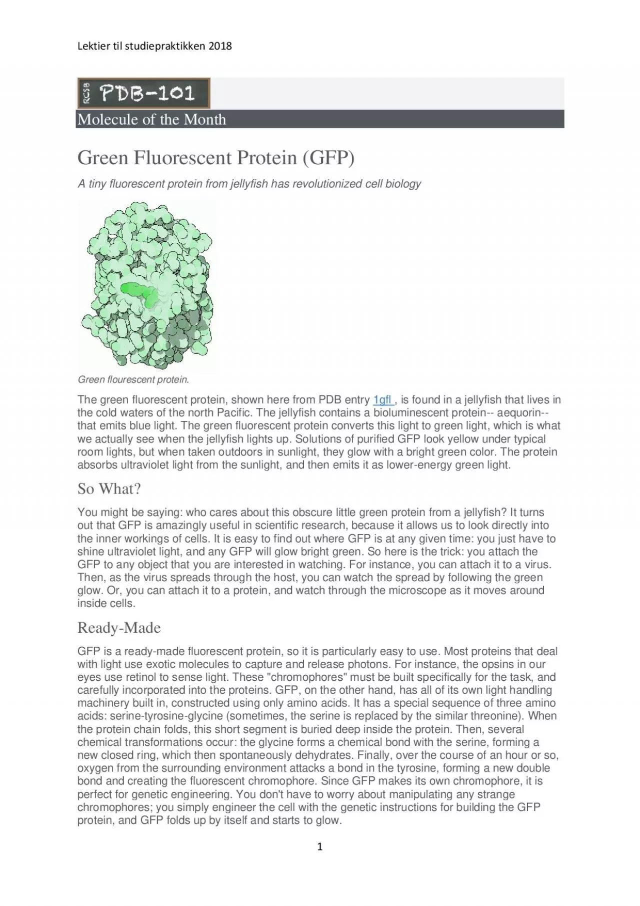
Lektier til studiepraktikken 2018 1 Molecule of the Month Green Fluorescent Protein (GFP) A tiny fluorescent protein from jellyfish has revolutionized cell biology Green flourescent protein. The green fluorescent protein, shown here from PDB entry 1gfl , is found in a jellyfish that lives in the cold waters of the north Pacific. The jellyfish contains a bioluminescent protein -- aequorin -- that emits blue light. The green fluorescent protein converts this light to green light, which is what we actually see when the jellyfish lights up. Solutions of purified GFP look yellow under typical room lights, but when taken outdoors in sunli ght, they glow with a bright green color. The protein absorbs ultraviolet light from the sunlight, and then emits it as lower - energy green light. So What? You might be saying: who cares about this obscure little green protein from a jellyfish? It turns out that GFP is amazingly useful in scientific research, because it allows us to look directly into the inner workings of cells. It is easy to find out where GFP is at any given time: you just have to shine ultraviolet light, and any GFP will glow bright gree n. So here is the trick: you attach the GFP to any object that you are interested in watching. For instance, you can attach it to a virus. Then, as the virus spreads through the host, you can watch the spread by following the green glow. Or, you can attach it to a protein, and watch through the microscope as it moves around inside cells. Ready - Made GFP is a ready - made fluorescent protein, so it is particularly easy to use. Most proteins that deal with light use exotic molecules to capture and release photon s. For instance, the opsins in our eyes use retinol to sense light. These "chromophores" must be built specifically for the task, and carefully incorporated into the proteins. GFP, on the other hand, has all of its own light handling machinery built in, co nstructed using only amino acids. It has a special sequence of three amino acids: serine - tyrosine - glycine (sometimes, the serine is replaced by the similar threonine). When the protein chain
folds, this short segment is buried deep inside the protein. Then , several chemical transformations occur: the glycine forms a chemical bond with the serine, forming a new closed ring, which then spontaneously dehydrates. Finally, over the course of an hour or so, oxygen from the surrounding environment attacks a bond i n the tyrosine, forming a new double bond and creating the fluorescent chromophore. Since GFP makes its own chromophore, it is perfect for genetic engineering. You don't have to worry about manipulating any strange chromophores; you simply engineer the cel l with the genetic instructions for building the GFP protein, and GFP folds up by itself and starts to glow. Lektier til studiepraktikken 2018 2 Engineering GFP The uses of GFP are also expanding into the world of art and commerce. Artist Eduardo Kac has created a fluorescent green rabbit b y engineering GFP into its cells. Breeders are exploring GFP as a way to create unique fluorescent plants and fishes. GFP has been added to rats, mice, frogs, flies, worms, and countless other living things. Of course, these engineered plants and animals a re still controversial, and are spurring important dialogue on the safety and morality of genetic engineering. Alba, the fluorescent bunny. Photo: Chrystelle Fontaine Improving GFP GFP is amazingly useful for studying living cells, and scientists are making it even more useful. They are engineering GFP molecules that fluoresce different colors. Scientists can now make blue fluorescent proteins, and yellow fluorescent proteins, and a host of others. The trick is to make small mutations that change the s tability of the chromophore. Thousands of different variants have been tried, and you can find several successes in the PDB. Scientists are also using GFP to create biosensors: molecular machines that sense the levels of ions or pH, and then report the res ults by fluorescing in characteristic ways. The molecule shown here (left) , from PDB entry 1kys , is a blue fluorescent protein that has been modified to sense the level of zinc ions. When zinc, shown here in red, binds to the mod
ified chromophore, shown here it bright blue, the protein fluoresces twice as brightly, creating a visible signal that is easily detected. Engineered biosensor for zinc io ns. Cytoskeleton staining with GFP and RFP in live HEKn cells ( https://www.thermofisher.com/dk/en/home/life - science/cell - analysis/cell - structure/celllight - ready - to - use - fluorescent - protein - based - reagents.html ) Lektier til studiepraktikken 2018 3 Exploring the Structure Green Fluorescent Protein The backbone of the entire protein is shown here on the left. The protein chain forms a cylindrical can (shown in blue), with one portion of the strand threading straight through the middle (shown in green). The chromophore is found right in the middle of the can, totally shielded from the surrounding environment. This shielding is essential for the fluorescence. The jostlin g water molecules would normally rob the chromophore of its energy once it absorbs a photon. But inside the protein, it is protected, releasing the energy instead as a slightly less energetic photon of light. The chromophore (shown in the close - up on the r ight) forms spontaneously from three amino acids in the protein chain: a glycine, a tyrosine and a threonine (or serine). Notice how the glycine and the threonine have formed a new bond, creating an unusual five - membered ring. References 1. 1kys: D.P. Baronde au, C.J. Kassmann, J.A. Tainer & E.D. Getzoff (2002) Structural chemistry of a green fluorescent protein Zn biosensor. Journal of the American Chemical Society 124, 3522 - 3524. 2. R.Y. Tsien (1998) The green fluorescent protein. Annual Review of Biochemistry 6 7, 509 - 544. 3. 1ema: M. Ormo, A.B. Cubitt, K. Kallio, L.A. Gross, R.Y. Tsien & S.J. Remington. Crystal structure of the Aequorea victoria green fluorescent protein. Science 273, 1392 - 1395. 4. 1gfl: F. Yang, L.G. Moss & G.N. Phillips (1996) The molecular structur e of green fluorescent proteins. Nature Biotechnology 14, 1246 - 1251. June 2003, David Goodsell doi: 10.2210/rcsb_pdb/mom_2003_6 RCSB PDB is a member