
Lecture 1 With DrHamzeh Al Share MD Original Slides for Prof Sameeh Al Saraireh Final 1 1 Fibrous proteins These proteins have a rod like structure They are not ID: 998536
Download Presentation The PPT/PDF document "Polypeptides and proteins -3" is the property of its rightful owner. Permission is granted to download and print the materials on this web site for personal, non-commercial use only, and to display it on your personal computer provided you do not modify the materials and that you retain all copyright notices contained in the materials. By downloading content from our website, you accept the terms of this agreement.


1. Polypeptides and proteins -3Lecture 1With Dr.Hamzeh Al-Shar`e , MDOriginal Slides for Prof. Sameeh Al-SarairehFinal1
2. 1- Fibrous proteins: These proteins have a rod like structure. They are not soluble in water. Collagen is an example these proteins often serve structural roles in cells. 2- Globular proteins: Due to their distribution of amino acids (hydrophobic inside, hydrophillic outside) they are very soluble in aqueous solutions (e.g Myoglobin) these proteins serve metabolic functions 3- Membrane proteins: Those membrane proteins that are embedded in the lipid bilayer have extensive hydrophobic amino acids that interact with the non-polar environment of the bilayer interior. Membrane proteins are not soluble in aqueous solutions.e.g:Rhodopsin Membrane proteins carry out transport activities, receptor functions, and other related processes Classification of Proteins Proteins can be classified based on their shape and solubility into three groups: 2
3. Fibrous Proteins1-Serve structural roles in cells- Fibrous proteins are often mechanically strong & highly cross-linked2-Insoluble in water 3-Secondary structure is simple based on one type only (Fibrous proteins have high alpha-helix or beta-sheet content) regardless the number 4-Functions in structure of the body or cell (tendons, bones, muscle, hair, skin)Most are structural proteins.Examples include:CollagenElastin KeratinFibroin General characteristics:Fibrous Proteins3
4. CollagenTensile strength results from the use of:The triple helix secondary structureThe assembly of tropocollagen subunits into a fibreChemical cross linking to strengthen the fibreIs the main structural protein of the various connective tissues in animals.The collagens are the most abundant proteins in the body. Make up from 25% to 35% of the whole-body protein contentThey occur in connective tissues where tensile strength is needed.Examples: skin, tendons, cartilage, bones.Fibrous Proteins4
5. Collagen is made up of three polypeptides (referred to as "α-chains") that are twisted around one another (tropocollagen) in a rope-like triple-helix and are held together by hydrogen bonds. Collagen is formed from tropocollagen subunits. The triple helix in tropocollagen is highly extended and strong. Features: (1) Three separate polypeptide chains arranged as a left-handed helix (each one is left-handed, but the tropocollagen is right-handed) - (note that an alpha-helix is right-handed).(2) 3.3 residues per turn (shortening occur due to presence of high number proline and glycine which cause kinking)(3) Each chain forms hydrogen bonds with the other twoFibrous Proteins5Structure of Collagen
6. Collagen type I (Most abundant)i) The fibers have diameter between 80 to 160nm. ii) Found in bone, dentin, skin, tendon, muscles and walls of blood vessels.Collagen type IIi) have a diameter <80nmii) found in invertiberal discs and hyaline cartilage.Collagen type III Found in spleen, muscle, and aorta.Collagen type IVFound around different types of the basement membranes and muscles. Collagen type VIt is found in embryonic cell cultures and the basement membranes.Collagen type VI It is found in muscle and skin.Types of collagen In humans at least there are 19 different collagens.Fibrous Proteins6
7. Types of collagen Type I collagen, which is the most common, is made up of: two identical peptide chains designated α1; (From the same gene)(b) one different chain designated α2 (Different gene)Both type II & III consist of three identical polypeptide chains. Compared to the α -helix, the collagen helix is much more extended. There are about 3.3 residues per turn of each of these helices due to kinkingFibrous Proteins7
8. Collagen Amino Acid CompositionMany modified amino acids are present: 4-hydroxyproline 3-hydroxyproline 5-hydroxylysine The structural features of collagen ranges from the amino acid sequence, tropocollagen molecules, collagen fibrils (consist of tropocollagens bounded together by cross-links) to collagen fibers.The hierarchical design of collagen.Nearly one residue out of three is Gly ( about 33% of total amino acids )Proline content is unusually highProline facilitates the formation of the helical conformation of each α-chain because its ring structure causes "kinks" in the peptide chain. Fibrous Proteins8
9. Connective tissues are proteins that support skin, bones, blood vessels and other organs.EDS usually affects your skin, joints and blood vessel walls. Ehlers-Danlos syndrome (EDS) EDS is a group of inherited connective tissue disorders, caused by a defect in the synthesis of collagen (Type I or III). The fragile skin and loose joints is often a result of abnormal genes that produce abnormal proteins that confer an inherited frailty of collagen (the normal protein "glue" of our tissues).Fibrous Proteins9There is no cure, and treatment is supportive, including close monitoring of the digestive, excretory and particularly the cardiovascular systems.
10. The type II and XI collagenopathies are a group of disorders that affect connective tissues. These disorders are caused by defects in type II or type XI collagen. Type II and type XI collagen disorders are grouped together because both types of collagen are components of the cartilage found in joints and the spinal column, the inner ear, and the jelly-like substance that fills the eyeballCausesMutations in the COL11A1, COL11A2, and COL2A1 genes cause collagenopathy, types II and XI. Note: Collagen 11 coding genes are COL11A1, COL11A2, and COL2A1 CollagenopathyFibrous Proteins10
11. Collagenopathy, type 2 alpha 1:refers to a wide range of conditions that can result from problems with cartilage collagen tissue due to a defect in the COL2A1 gene. Defects in the COL2A1 gene result in defective or reduced collagen production which in turn affects the development of connective tissues including bones. Defects in COL2A1 gene affect production of collagen Type 2 and Type 11 .Fibrous Proteins11
12. 12Symptoms of Collagenopathy, type 2 alpha 1Abnormal bone developmentShort statureEnlarged jointsCurved spinePremature arthritisVision problemsHearing problemsCleft palateSmall lower jawVarious facial anomalies
13. KeratinCharacteristicsTough and insoluble in waterMain constituent of hair, nails and tooth enamel Two major conformational groups (a) alpha-keratin whose peptide backbone forms a α-helix(b) β-keratin whose backbone forms a β-sheet structure.Keratin is the key structural material making up the outer layer of human skin.Fibrous Proteins13
14. α-KeratinMade up of α-helix alpha-keratin is found in hair, nails, outer layer of skin. It forms almost the entire dry weight of these materials. The entire secondary structure is a dimer of two alpha-helices.It is rich in amino acids that favours alpha-helix formationThese hydrophobic side chains are on the alpha-helix surface-explaining its insolubility.It is also rich in Cys residues. Two Cys residues form disulphide bridges in alpha-keratin, and link the alpha-helices together. The more disulphides, the stronger the alpha-keratin.Fibrous Proteins14
15. Motor ProteinsThe most prominent example of a motor protein is the muscle protein myosin and actin which performs the contraction of muscle fibers in animals. - Sarcomer is the smallest functional structure of the muscleFibrous Proteins15
16. - Sarcomere is made up of three different filament:1- Myosin the thick filaments2- Actin the thin filaments. 3- Proteins that stabilize the positions of the thick and thin filaments, and proteins that regulate the interactions between thick and thin filaments.= The myosin head attaches to an actin filament within the sarcomere of a myofibril then pull towards the centre of the sarcomere. In the process, the sarcomere length shortens and the muscle contracts. ATP needed to dissociate myosin head rom actinMotor Proteins
17. Myosins are motor proteins that interact with actin thin filament and hydrolyse ATP to generate movement. Myosin is a hexamer that consists of two heavy chains (220 kDa), and four light chains (~ 20 kDa each) paired into two regulatory light chains and two essential light chains. Myosin structureFibrous Proteins17Motor Proteins
18. 18The globular head (S1) forms the actin binding site and the ATPase siteS1 can be further divided into 3 subdomains: 1-the N-terminal nucleotide (ATP) binding domain 2-the central actin binding domain 3-the C-terminal actin binding domain The heavy chain consists of three proteolitically defined domains: 1- subfragment 1 (S1) (Contain actin-binding site & ATPase ) 2- subfragment 2 (S2) 3- light meromyosin (LMM). Myosin structure
19. Actin is a 42 kDa adenine nucleotide-binding protein that made of 375 amino acids, and it is essential for so many cell functions. Actin is found in two major forms: Globular which is the monomeric form (G-actin) that can spontaneously polymerise into the filamentous form (F-actin) at physiological salt concentration. Actin Actin monomers are arranged in a two-strand helix. Fibrous Proteins19
20. Tropomyosin (Tm) is a right-handed helical protein which forms a coiled dimer that cooperatively binds with actin thin filament. Each Tm chain is composed of 284 amino acids, and a tight hydrophobic interaction between two chains holds them together Tropomyosin is always found associated with actin. Each tropomyosin spans the length of seven actin monomers. Tropomyosin (Tm) Fibrous Proteins20
21. Troponin is the calcium-based regulator of striated muscle contraction. Troponin is a heterotrimeric complex that is composed of three interacting subunits: 1- Troponin C which is the Calcium sensor subunit,2- Troponin I which is the Inhibitory subunit and 3- Troponin T which is the Tropomyosin binding subunit. Troponin complex (Tn)A schematic representation of the interaction between the troponin complex and the rest of the thin filament. The black arrows indicate the interaction between actin-tropomyosin and troponin in the presence and absence of calcium Fibrous Proteins21
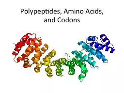
22. Globular proteins, also known as sphero-proteins, are proteins formed by compacted amino acid chains, which are folded into intricate shapes that often roughly resemble spheres. Globular Proteins A key difference between globular proteinsand fibrous proteins is that the former type of protein is usually soluble in water, while the latter type is not. Globular proteins include enzymes, transport proteins, regulatory proteins, proteins with many other functions. Globular proteins comprise the most varied type of proteins. Globular proteins are soluble in aqueous solution. To achieve this, globular proteins generally have polar residues on the surface and hydrophobic residues on the interior.22
23. All alpha: Proteins that contain only alpha helical secondary structure. Myoglobin is an example of an all alpha protein.All beta: Protein that contain only beta-sheet secondary structure. Tenascin is an example of an all beta protein.Alpha/beta: Proteins that contain alternating alpha-helical and beta-sheet secondary structure elements. Triose Phosphate Isomerase is an example of an alpha/beta protein. Alpha + Beta: In these proteins the alpha helical and beta sheet regions occur in independent regions of the molecule. Ribonuclease A is an example of an alpha+beta protein.Classification of Globular Proteins According to Secondary StructureGlobular Proteins23
24. Oxygen-binding proteinsMyoglobin and Hemoglobin are the two oxygen-binding proteins present in large multicellular organisms.Myoglobin stores the oxygen in the muscles.Hemoglobin transports oxygen in the blood and is located in the red blood cellsGlobular Proteins24
25. Myoglobin (Mb) Three critical functions for Mb: 1- it holds the heme group, 2- it protects the heme iron from oxidation, 3- it provides a pocket into which the O2 can fit.75% of structure is -helix in 8 regions, these are termed helices A, B, C, D, E, F, G, and H.Myoglobin consists of a single polypeptide chain of 153 amino acids attached to a single heme groupMyoglobin interior almost entirely non-polar residues Mb Stores and facilitates oxygen diffusion in muscles especially in heart and skeletal muscle. The eight α helical segments are folded into a globular structure, creating a cradle (box) and within this cradle lies a single heme group and the binding site of O2. The heme of myoglobin lies between helices E and F.Globular Proteins25
26. Heme is a complex of porphyrin and ferrous iron (Fe2+). Porphyrins are a group of organic compound that have four pyrrole subunits interconnected via α-methylene bridges (=CH-)Structure of heme in myoglobinThe hydrophobic environment in the interior of myoglobin or hemoglobin prevents the oxidation of iron.Globular Proteins26
27. Heme structureThe iron is held in the center of the porphyrin ring. Iron ions prefer to interact with six ligands. Four of the ligands to this iron ion are provided by nitrogen atoms in the pyrrole ring system. The fifth ligand is provided by a nitrogen atom from the imidazole group of His 93 (proximal histidine) (also known as His F8 which is the eighth residue of the 'F helix' of myoglobin). The sixth ligand to iron is provided by molecular oxygen, which binds to the heme group in a pocket formed by Mb. Globular Proteins27