
Older Brain Structures The Brainstem is the oldest part of the brain beginning where the spinal cord swells and enters the skull It is responsible for automatic survival functions 2 Brainstem ID: 784566
Download The PPT/PDF document "The Brain Module 4 1 The Brain:" is the property of its rightful owner. Permission is granted to download and print the materials on this web site for personal, non-commercial use only, and to display it on your personal computer provided you do not modify the materials and that you retain all copyright notices contained in the materials. By downloading content from our website, you accept the terms of this agreement.

Slide1
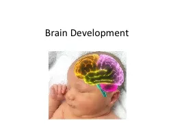
The BrainModule 4
1
Slide2The Brain: Older Brain StructuresThe Brainstem
is
the oldest part of the brain, beginning where the spinal cord swells and enters the skull. It is responsible for automatic survival functions.
2
Slide3BrainstemThe Medulla [muh-DUL-uh] is the base of the brainstem that controls heartbeat and breathing.
3
Slide4Brainstem--PONSAppears just above the medullaHelps coordinate movementsHouses nerve circuits that regulate sleep and dreaming cycle.Also acts as the “bridge” that connects the brain stem to the cerebellum.4
Slide5BrainstemThe Thalamus [THAL-uh-muss] is the brain’s sensory switchboard, located on top of the brainstem. It directs messages to the sensory areas in the cortex and transmits replies to the cerebellum and medulla.
5
Slide66Brainstem
Reticular Formation
is
a nerve network in the brainstem that plays an important role in controlling arousal.
Slide7CerebellumThe “little brain” attached to the rear of the brainstem. It helps coordinate voluntary movements and balance.
Slide8The BrainA brain lesion experimentally destroys brain tissue to study animal behaviors after such destruction.
8
Techniques to Study the Brain
Hubel (1990)
Slide9Clinical ObservationClinical observations have shed light on a number of brain disorders. Alterations in brain morphology due to neurological and psychiatric diseases are now being catalogued.9
Tom Landers/ Boston Globe
Slide10Electroencephalogram (EEG)An amplified recording of the electrical waves sweeping across the brain’s surface, measured by electrodes placed on the scalp.
10
AJ Photo/ Photo Researchers, Inc.
Slide11PET Scan11PET (positron emission tomography) Scan is a visual display of brain activity that detects a radioactive form of glucose while the brain performs a given task.
Courtesy of National Brookhaven National Laboratories
Slide12MRI Scan12MRI (magnetic resonance imaging)
uses magnetic fields and radio waves to produce computer-generated images that distinguish among different types of brain tissue. Top images show ventricular enlargement in a schizophrenic patient. Bottom image shows brain regions when a participants lies
.
fMRI
-reveals function as well as structure
.
https://
aeon.co/videos/contestants-have-five-minutes-in-an-fmri-to-love-someone-as-hard-as-they-can
Both photos from Daniel Weinberger, M.D., CBDB, NIMH
James
Salzano
/
Salzano
Photo
Lucy Reading/ Lucy Illustrations
Slide13The Limbic SystemThe Limbic System is
a doughnut-shaped system of neural structures at the border of the brainstem and cerebrum, associated with emotions such as fear, aggression and drives for food and sex. It includes the hippocampus, amygdala, and hypothalamus.
13
Slide14AmygdalaThe Amygdala [ah-MIG-dah-la] consists of two lima bean-sized neural clusters linked to the emotions of fear and anger.
14
Slide15HypothalamusThe Hypothalamus lies below (hypo
) the thalamus. It directs several maintenance activities like eating, drinking, body temperature, and control of emotions. It helps govern the endocrine system via the pituitary gland.
Governs the 4F’s
15
Slide16Lateral hypothalamus: “Lunch!”(Involved in “hunger messages”)
V
entromedial hypothalamus: “
V
omit!!!”
(The “satiety center”; “You’ve had enough!”)
16
Slide17Reward CenterRats cross an electrified grid for self-stimulation when electrodes are placed in the reward (hypothalamus) center (top picture). When the limbic system is manipulated, a rat will navigate fields or climb up a tree (bottom picture).17
Sanjiv Talwar, SUNY Downstate
Slide18HippocampusProcesses memoryConnects your present to your past and remember the location of things in space.18
Slide19The Cerebral CortexThe intricate fabric of interconnected neural cells that covers the cerebral hemispheres. It is the body’s ultimate control and information processing center.
19
Slide20Structure of the CortexEach brain hemisphere is divided into four lobes that are separated by prominent fissures. These lobes are the frontal lobe
(forehead),
parietal lobe
(top to rear head),
occipital lobe (back head) and
temporal lobe
(side of head).
Slide21Functions of the CortexThe Motor Cortex is the area at the rear of the frontal lobes that control voluntary movements. The Sensory Cortex
(parietal cortex) receives information from skin surface and sense organs.
21
Slide22Frontal LobesHigher Mental functionsIncludes the motor CortexPre-frontal Cortex—decision-making and reasoning skillsPhineas Gage.http://learner.org/vod/vod_window.html?pid=159222
https://
youtu.be/UFHMKRElkSY
Slide23Parietal LobesSensory (somatosensory) CortexRight Lobe: helps keep track of your body parts.Left Lobe: specialized in locating the source of speech sounds, as when someone calls your name.It also works with the temporal lobe to extract meaning from speech, and writing.23
Slide24Occipital LobeYou have eyes in the back of your head!!Receives stimulation relayed from the eyes to the visual cortex, which constructs our moving picture of the outside world.24
Slide25Temporal LobesAuditory CortexHelps make sense of sounds.25
Slide26Visual FunctionThe functional MRI scan shows the visual cortex is active as the subject looks at faces.
26
Courtesy of V.P. Clark, K. Keill, J. Ma. Maisog, S. Courtney, L.G.
Ungerleider, and J.V. Haxby,
National Institute of Mental Health
Slide27Auditory FunctionThe functional MRI scan shows the auditory cortex is active in patients who hallucinate.
27
Slide28Language28
Aphasia
is an impairment of language, usually caused by left hemisphere damage either to
Broca’s
area
(impaired speaking) or to
Wernicke’s area
(impaired understanding
) or in the
Angular Gyrus
that leaves people unable to read aloud.
Slide29Specialization & Integration
Brain activity when hearing, seeing, and speaking words
29
Slide30The Brain’s Plasticity
The
brain is sculpted by our genes but also by our experiences.
Plasticity
refers to the brain’s ability to modify itself after some types of injury or illness
.
Our Brains are more plastic when we are children
Constraint-Induced Therapy
Neurogenesis: formation of new
neurons
https://
www.youtube.com/watch?v=VaDlLD97CLM-
Brain Plasticity the story of Jody
https://
www.ted.com/talks/sandrine_thuret_you_can_grow_new_brain_cells_here_s_how
30
Slide31Our Divided BrainOur brain is divided into two hemispheres.
The left hemisphere processes reading, writing, speaking, mathematics, and comprehension skills. In the 1960s, it was termed as the dominant brain
.
31
Slide32Splitting the BrainA procedure in which the two hemispheres of the brain are isolated by cutting the connecting fibers (mainly those of the corpus callosum) between them
.
*band of fibers that connect/carry the messages between the hemispheres.
32
Corpus Callosum
Martin M. Rother
Courtesy of Terence Williams, University of Iowa
Slide33Split Brain PatientsWith the corpus callosum severed, objects (apple) presented in the right visual field can be named. Objects (pencil) in the left visual field cannot.https://www.youtube.com/watch?v=lfGwsAdS9Dc
33
Slide34Divided Consciousness34
Slide35Try This!35
Try drawing one shape with your left hand and one with your right hand,
simultaneously
https
://www.nobelprize.org/educational/medicine/split-brain
/
BBC
Slide36Non-Split Brains36People with intact brains also show left-right hemispheric differences in mental abilities.
A number of brain scan studies show normal individuals engage their right brain when completing a perceptual task and their left brain when carrying out a linguistic task.