
of specialized cells and cell products organized to perform one or more select functions Histology study of tissues All cells in the body are classified into one of the four tissue types Epithelial ID: 1037266
Download Presentation The PPT/PDF document "119 Tissues are collections" is the property of its rightful owner. Permission is granted to download and print the materials on this web site for personal, non-commercial use only, and to display it on your personal computer provided you do not modify the materials and that you retain all copyright notices contained in the materials. By downloading content from our website, you accept the terms of this agreement.


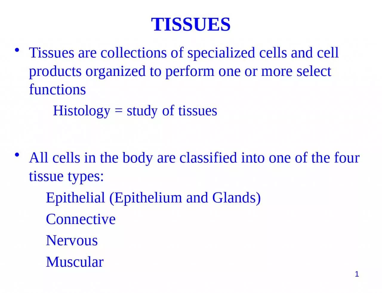
1. 119Tissues are collections of specialized cells and cell products organized to perform one or more select functionsHistology = study of tissuesAll cells in the body are classified into one of the four tissue types: Epithelial (Epithelium and Glands) Connective Nervous MuscularTISSUES
2. 120Epithelial Tissue: our first tissue typeTypes of Epithelial Tissue1. Epithelial sheets (“epithelium”)2. Glands
3. Epithelial TissueLecture OutlineCharacteristics of Epithelial SheetsCell-Cell Junctions : Gap Junctions, Tight Junctions, Desmosomes, and Lateral InterdigitationsNaming Epithelial Sheets by Thickness and Cell shapeGlands : Endocrine and Exocrine121
4. 122Locations: covers body surface (outermost part of skin) lines all hollow structures in body Functions:Physical protectionControl permeability Produce specialized secretionsEpithelium: a layer of tightly adjoining related by their embryology and function
5. 123Characteristics of Epithelial sheets:Cells are polarizedLittle space between cellsCells tightly held together by junctionsAvascular (no blood vessels between cells)Specialized for absorption or protectionContinuous rate of cell division
6. 124Epithelial Cells have Polarity (sided-ness)Apical (free or exposed surface)Basal (surface anchored to connective tissue)Lateral(neighboring epithelial cells)
7. 125Epithelial cells are attached at the basal surface to a basement membraneThe epithelial cells are attached to connective tissue through the glue-like proteins of the basement membrane. We sometimes use the phrases “basal lamina” and “basement membrane” interchangeably
8. 126The avascular conceptLittle space between adjacent cells for blood vessels.Epithelial cells are usually kept alive by diffusion of oxygen and nutrients from the blood vessels in the adjacent connective tissue below the basal laminaEpithelial layers are limited in thickness – diffusion is poor over long distances or dense areasEpithelium one layer in thicknessEpithelium multiple layers in thickness
9. 127Some Epithelial Cells may have specializations on their apical surfacesMICROVILLISmall fingerlike projections of the apical or basal surface of an epithelial cell(may be hundreds per cell)Microvilli increase surface area for the cell
10. 128Some Epithelial Cells have specializations on their apical surfacesCILIALong slender extensions of the apical cell surfaceCilia have a core of microtubulesCilia exhibit rhythmical movement
11. 129cilia
12. 130Epithelial Cells are tightly held to their neighboring epithelial cellsCells attach via cell adhesion moleculesCells attach at specialized cell junctions:Gap junctions, Desmosomes, Tight junctions, Lateral Interdigitations
13. 131Connections between Epithelial Cells : Gap JunctionsCells are connected by membrane proteins that form pores or channels between cellsCytoplasm of one cell is continuous with cytoplasm of adjacent cell allowing ions to move between cellsImportant in electrical signaling and organs where cells work in close synchrony
14. 132Connections between Epithelial Cells : Tight JunctionsThe plasma membranesof adjacent cells are “fused” together bymembrane proteins
15. 133Connections between Epithelial Cells : Tight JunctionsTight junctions form an occluded zone just under the apical surface where the neighboring cells have fusedtheir membranes togetherExtends all the way aroundthe cell, with different neighborsWater and solutes cannot pass between cellsApical surface
16. 134Connections between Epithelial Cells : DesmosomesAdjacent cellsjoined by membrane proteinsand a glue-likematerial
17. 135Review: Tight Junctions, Gap Junctions, and Desmosomes and their relative locations
18. 136Lateral interdigitations Adjacent cells have intertwined membranesTight JunctionDesmosomeLateral Interdigitation
19. 137Basement membrane joins epithelial layer to underlying connective tissue layerEpithelial stem cells replace short-lived epithelial cellsBecause epithelial sheets are in vulnerable body positions, they have a high rate of cell division to constantly replace damaged cellsRemember that carcinomas, tumors of epithelial cells, account for 90% of all cancers.Other features of typical epithelium
20. 138Number of cell layersSimpleStratifiedShape of apical surface cellsSquamous Cuboidal ColumnarEpithelial sheets are described with 2 adjectives One cell thickMore than one cell thickFlat, broad cellsCube-shapedTall, narrow cells
21. 139Simple Squamous Epithelium Single layer of flat cells; little protection, allows for fast diffusion Locations: lines all the cardiovascular organs (heart, vessels), air spaces in lungs, etc.
22. 140Simple Cuboidal EpitheliumOne layer thick; cells block–like in shape (as tall as they are wide)Function: allows for regulated exchange; cytoplasm typically holds many mitochondria to drive active transport processes; may have microvilliLocations: kidneys, ducts
23. 141 Simple Columnar EpitheliumCells taller than they are wide, one layer thick; cells often have microvilli or cilia on apical surfaceFunctions: Some protection; active transport; often have mucous cells interspersedLocations: intestines
24. Stratified Squamous EpitheliumMany layers of cells thick apical surface is flattened grows from basal surfaceFunctions: Mainly as a barrier - denies access to connective tissue below; does not allow for exchange through epitheliumLocations: surface of skin, lining of mouth, anus, vagina, etc.142
25. 143Transitional EpitheliumUnique to urinary system
26. Ciliated Pseudostratified Columnar Epithelium the special epithelium of the respiratory systemThe ciliated surface of the respiratory tract144
27. 145Endocrine glandsRelease hormones into surrounding fluid to be picked up and carried away by the bloodExocrine glandsSecrete through ducts onto the surface of the organGlandular epitheliacells of epithelial line which are specialized to produce and secrete materials outside the cellMerocrine (product released through exocytosis)Apocrine (involves the loss of both product and cytoplasm)Holocrine (destroys the cell)
28. 146The Structure of Endocrine GlandsProduct is released from cell into extracellular fluidProduct is picked up and carried away by bloodstream where it may affect distant tissues
29. 147Mechanisms of Exocrine Gland Secretion: Merocrine Product is packaged into secretory granulesReleased from cell into duct by exocytosisExample: salivary glands, pancreas
30. 148Mechanisms of Exocrine Gland SecretionApocrineApical portion of cell is shed, taking product with it into ductExample: mammary glands
31. 149Mechanisms of Exocrine Gland Secretion: HolocrineThe entire cell bursts, releasing product into duct. Cell dies and must be replacedExample: oil glands associated with hair follicles
32. 150Individual secretory cells are embedded within an epithelium (example: Goblet cells) Structure of Unicellular Glands
33. 151Structural Classification of Multicellular Glands Don’t memorize these terms, just appreciate the different levels of complexity