
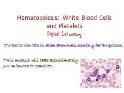
es monocytes and their benign disorders The white blood cells leucocytes may be divided into two broad groups 1the phagocytes Granulocytes which include three types of cell neutrophils ID: 920324
Download Presentation The PPT/PDF document "The white cells 1: granulocy" is the property of its rightful owner. Permission is granted to download and print the materials on this web site for personal, non-commercial use only, and to display it on your personal computer provided you do not modify the materials and that you retain all copyright notices contained in the materials. By downloading content from our website, you accept the terms of this agreement.


Slide1
The white cells 1: granulocy es,monocytes and their benigndisorders
Slide2The white blood cells (leucocytes) may be divided into two broad groups:1-the phagocytes Granulocytes, which include three
types of cell-
neutrophils
(polymorphs),
eosinophils
and
basophils
-together with
monocytes
comprise the phagocyte
2-immunocytes
Note : Only
mature
phagocytic
cells and lymphocytes are found in
normal peripheral blood
Slide3Slide4GranulopoiesisThe blood granulocytes and monocytes are formedin the bone marrow from a common precursor cell
In the
granulopoietic
series progenitor
cells which are:
myeloblasts
,
promyelocytes
and
myelocytes
form a
proliferative or mitotic pool
of cells
while
the
metamyelocytes
, band and segmented granulocytes
make up
a post-mitotic maturation
compartment
Large
numbers of band and segmented
neutrophils
are held in the marrow as a
'reserve
pool' or storage
compartment
.
Slide5Slide6In the bone marrow
normally
contains more myeloid cells than
erythroid
cells in the ratio of 2 : 1 to 12 :
1
the
largest proportion
being
neutrophils
and
metamyelocytes
.
In the stable or normal state, the bone
marrow storage
compartment contains 10-15 times
the number
of granulocytes
found
in the
peripheral blood
.
Following
their release from the bone marrow,
granulocytes spend only 6-10 h in the circulation
before moving into the tissues where they
perform their
phagocytic
function
Slide7In the blood stream
there
are two pools usually of about equal
size
:
the
circulating pool
(included in the
blood count
)
and
the
marginating
pool
(not included in
the blood count).
It
has been estimated that they
spend on average 4-5 days in the tissues before they
are destroyed during defensive action or as the
result of senescence.
Slide8Control of granulopoiesis: myeloid growth factors
Many growth factors are involved in this
maturation process including
:
interleukin-1
(IL-1
)
IL-3
IL-5 (for
eosinophils
),
IL-6
IL-11
granulocytemacrophage
colony stimulating
factor (GM-CSF
)
granulocyte
CSF (G-CSF)
monocyte
CSF (MCSF
)
Slide9Function of growth factors The growth factors stimulate proliferation and
differentiation
and also affect
the
function
of the mature cells on which they act
e.g.
Increased granulocyte and
monocyte
production in
response to an infection is induced by
increased production
of growth factors from
stromal
cells
and T
lymphocytes, stimulated by
endotoxin
, IL-1
or
tumour
necrosis factor (TNF)
Slide10Slide11The normal function of neutrophils and
monocytes
may
be divided into
three phases:
Chemotaxis
(cell mobilization and migration)
The
phagocyte
is attracted to bacteria or the site
of inflammation
by
chemotactic
substances.
Phagocytosis
The foreign material (e.g. bacteria,
fungi
) or dead or damaged cells of the host are
Phagocytosed
Killing and digestion
This occurs by
oxygen dependentand oxygen-independent pathways.
Slide12Functional Disorders of Phagocytic Leucocytes12
Slide13Disorders characterised by neutrophil dysfunction
1. Impaired adhesion:
Congenital leucocyte adherence deficiency (Deficiency of CD11/CD18 surface glycoproteins)
Drugs: corticosteroids, alcohol
2. Impaired motility:
Hyperimmunoglobulin
E syndrome
Chediak
-Higashi syndrome
Diabetes mellitus, Hodgkin’s disease, leprosy
3. Impaired
microbicidal
killing:
Chronic granulomatous disease
Myeloperoxidase deficiency
Chediak
-Higashi syndrome
Leukaemias
13
Slide14Benign disordersof granulocyte morphology
1-Pelger-Huet
anomaly
In this
uncommon condition
bilobed
neutrophils
are found in the peripheral
blood. Occasional
unsegmented
neutrophils
are also seen.
Inheritance is
autosomal
dominant.
Slide152-May-Hegglin anomaly In this rare condition the
neutrophils
contain basophilic inclusions of RNA
in
the cytoplasm
.
There is
an associated mild thrombocytopenia with
giant platelets
.
Inheritance
is
autosomal
dominant.
Slide163-The Chediak-Higashi syndrome is inherited
in an
autosomal
recessive
manner,
and there are
giant granules in the
neutrophils
,
eosinophils,monocytes
and lymphocytes accompanied by
neutropenia
, thrombocytopenia
and
marked
hepatosplenomegaly
.
disorders may be associated with severe
disease.
Slide17Common morphological abnormalities
Hypersegmented
forms occur in
megaloblastic
Anaemia
Dahle
bodies and toxic changes
in infection.
Slide18The 'drumstick' appears on the nucleus ofa proportion of the neutrophils in normal females
and is caused by the presence of two X chromosomes.
Pelger
cells
are seen in the benign congenital
abnormality but also in patients with acute myeloid
leukaemia
or
myelodysplasia
Slide19Shift to the left:
increase in the number of band forms and the occasional presence of more primitive cells in the peripheral blood.
Leukoerythroblastic
picture:
a presence of a shift to left plus nucleated RBC in the peripheral blood.
Slide20Slide21Slide22Causes of leucoerythroblasticblood film
Metastatic neoplasm in the marrow
Primary
myelofi
brosis
Acute
and
chronic
myeloid
leukaemia
Myeloma, lymphoma
Miliary
tuberculosis
Severe
megaloblastic
anaemia
Severe
hemolysis
Osteopetrosis
22
Slide23Causes of leucoerythroblasticblood film
Metastatic neoplasm in the marrow
Primary
myelofi
brosis
Acute
and
chronic
myeloid
leukaemia
Myeloma, lymphoma
Miliary
tuberculosis
Severe
megaloblastic
anaemia
Severe
hemolysis
Osteopetrosis
23
Slide24Slide25The Leukomoid Reaction
a
reactive excessive leukocytosis with the presence
of immature cells (
myeloblasts
,
promyelocytes
and
myelocyte
) in the peripheral blood.
In
other words (
leukocytosis + left shift
)
. Usually
it is a neutrophil leukocytosis
. It can be lymphoid
Causes :
Severe infection
Severe
haemolysis
Metastatic cancer
Slide26Slide27