
Finding or Clinical Pathology Alan C Yeung MD Li Ka Shing Professor of Medicine Chief Div of Cardiovascular Medicine Director Cardiovascular Health Stanford University Myocardial Bridges Pr ID: 946992
Download Pdf The PPT/PDF document "Myocardial Bridge Incidental" is the property of its rightful owner. Permission is granted to download and print the materials on this web site for personal, non-commercial use only, and to display it on your personal computer provided you do not modify the materials and that you retain all copyright notices contained in the materials. By downloading content from our website, you accept the terms of this agreement.


Myocardial Bridge: Incidental Finding or Clinical Pathology? Alan C. Yeung, MD Li Ka Shing Professor of Medicine Chief, Div of Cardiovascular Medicine Director, Cardiovascular Health Stanford University Myocardial Bridges Present in 30 - 80% of populat
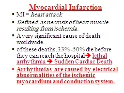
ion by autopsy (by angiography) Occurs in ~40% of patients with angina and normal coronary arteries Most common in the LAD Generally considered benign, but have been associated with myocardial ischemia/ infarcation , VT, and sudden death Alegria et al. E
ur Heart J 2005;26:1159 - 1168 Presentation Symptoms typically do not develop before the third decade Patients typically have exertional chest pain, although CP may occur with mental stress. Over time, symptoms seem to become more persistent Patients ofte
n have a lot of PVCs, and VT/syncope can be a presenting symptom Reports of anteroseptal ischemia on nuclear perfusion scans, septal ischemia/infarction on MRI and autopsy Recently by stress echo, we have found a focal mid septal “buckling” Focal mid septal
“buckling” Occurs end - systole/early diastole with apical sparing Lin et al. J Am Heart Assoc 2013;2:e000097 Myocardial Bridging - Anatomy Echo - lucent half moon sign (halo) felt to be pathognomonic, although not 100% sensitive ≥10% systolic c
ompression Normal LAD IVUS 57% positive Lin et al. J Am Heart Assoc 2013;2:e000097 Myocardial Bridging - Pressure FFR with adenosine not sensitive enough for detecting ischemia with myocardial bridging — may improve sensitivity by diastolic FFR with dobutami
ne Escaned et al. J Am Coll Cardiol 2003;42:226 - 33 Ischemia Within Bridge Assumption has been that ischemia is distal to the myocardial bridge We hypothesized that the ischemia occurs within the bridge, rather than distal to it Studied ~60 patients with
IVUS, as well as combination pressure and Doppler flow velocity proximal to, within, and distal to the bridge at baseline and with dobutamine stress Reported first 18 patients (age 16 to 62 years, median 43 years) Lin et al. J Am Heart Assoc 2013;2:e000097 Baseline
Pressure and Flow Pressure and Flow at Stress dFFR=0.74 dFFR=0.88 Significant dFFR Within Bridge All had significantly abnormal dFFRs The patients with the abnormal distal dFFR notably had the longest MBs (mean 40.5mm) and/or had 2 MBs With rest and
stress, the peak diastolic flow velocities within the bridge were significantly higher than those proximally or distally Ischemia Within Bridge due to Venturi Effect Venturi effect: moving through a narrowed area, velocity must increases (principle of continuity)
with a required drop in pressure (conservation of energy by Bernoulli’s equation) The narrowest lumen within a bridge is at end - systole/early diastole Conclude that ischemia is local to the MB rather than distal to it (ischemia within septal branches) As
sociate with findings on stress echo of focal mid septal buckling Stanford Is Myocardial Bridging truly benign? Impact of myocardial bridging induced arterial compression on atherosclerotic plaque formation Ryotaro Yamada, MD, PhD; Ingela Schnittger, MD; Jennifer
A. Tremmel, MD; Shin Lin, MD, PhD; Paul G. Yock, MD; Peter J. Fitzgerald, MD, PhD; Yasuhiro Honda. MD Division of Cardiovascular Medicine Stanford University Medical Center, Stanford, CA Stanford Cx LAD IVUS Parameters Max PB prox Plaque burden (PB)
= (EEM - CSA – Lumen CSA) / EEM - CSA Up to 20 mm proximal from MB entrance Max PB MB within MB MB segment 20 mm Proximal ref Length from LAD ostium to MB Halo thickness EEM - CSA (Sys & Dia ) & Arterial compression Branch within MB MB length
Stanford Proximal ref MB segment Max PB in Proximal vs. MB segment 0 20 40 60 80 100 p Max PB (%) Stanford Arterial Compression and Max PB prox Younger adults (age ≤ 53 years ) with ≤ one risk factor 0 20 40 60 80 100 0 20 40 60 Arterial compression (%
) r=0.565, p1 Max PB prox (%) Stanford • Max PB prox was significantly greater than Max PB MB . • Arterial compression had a significant positive correlation to Max PB prox , but not to Max PB MB . • No other IVUS properties of MB correlated with
Max PB prox . • In multivariate analysis, arterial compression was independently associated with Max PB prox . • When isolated from the influence of age and coronary risk factors, the correlation between arterial compression and Max PB prox showed an even st
ronger relationship. Summary Ms. S. K. • December 2012 : 52 years old previously healthy woman admitted to OSH with NSTEMI and troponin of 0.8 with no ECG changes. • January 2013 : Coronary angiogram showed no significant CAD. Mid LAD myocardial bridge.
Ms. S. K. 1. Early February 2013 : Admitted with recurrent severe chest pain. Second cor. a ngiogram showed rapid progression of CAD in one month, suggestive of plaque rupture. 2. IVUS showed 41 mm long MB, halo thickness of 1.0mm. Maximal systolic
compression was 22 % ( 2.98mm 2 /3.55mm 2 ) Ms . S.K. Angiogram II 60% CAD MB January 2013: Angiogram I February 2013: Angiogram II Conclusions Myocardial bridges are common, but not completely benign Coronary angiography rarely identifies th
em, IVUS is needed (stress echo and CTA can also be helpful) Hemodynamic assessment of symptomatic bridges shows an increase in flow velocity and a decrease in pressure ( dFFR ) within the bridge more so than distal to it, suggesting a local ischemic effect (i.e. septal
ischemia). S uch an assessment may be helpful in identifying hemodynamically significant bridges in patients with angina and normal appearing coronary arteries Plaque burden is increased in the proximal reference segment. Whether these plaques have increased vuln