
What we hope to learn The basic building blocks amino acids Secondary structure Forces that drive folding Motifs or supersecondary structure Domains Finding out more about structures ID: 1033929
Download Presentation The PPT/PDF document "Visualizing Protein Structures" is the property of its rightful owner. Permission is granted to download and print the materials on this web site for personal, non-commercial use only, and to display it on your personal computer provided you do not modify the materials and that you retain all copyright notices contained in the materials. By downloading content from our website, you accept the terms of this agreement.


1. Visualizing Protein Structures What we hope to learn The basic building blocks: amino acids Secondary structure Forces that drive folding Motifs or supersecondary structure Domains Finding out more about structures How to visualize molecules with VMD How to edit Protein Data Bank filesCore
2. Why we should care more about structure and less about informatics…
3. Why we should care more about structure and less about informatics…Structure and dynamics are needed to infer function.
4. Why we should care more about structure and less about informatics…Structure and dynamics are needed to infer function.There is more to bioinformatics than AGCT or protein sequence
5. Why we should care more about structure and less about informatics…Structure and dynamics are needed to infer function.There is more to bioinformatics than AGCT or protein sequenceRNA is more important than DNA
6. “The known biological functions of RNA continue to grow in number and to expand in scope. RNA has been transformed from a molecule with a minor role in protein synthesis, to an important player in all of molecular biology.”“Furthermore, it has been shown that stem-loops in the mRNA can bind to the protein product of the message to regulate its synthesis, pseudoknots in retroviral mRMAs cause programmed frameshifts that produce the correct ratios of proteins required for viral propagation.”Tinoco, “How RNA folds.” J Mol Biol. 1999 Oct 22;293(2):271-81. Review. Ribozymes, Riboswitches, RNAi
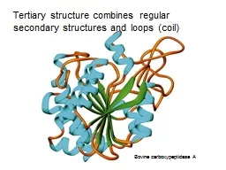
7. From: Brandon & Tooze, “Introduction to Protein Structure”primary (1º) secondary (2º) tertiary (3º) quaternary (4º) (from Mount, “Bioinformatics Sequence and Genome Analysis”
8. From: Brandon & Tooze, “Introduction to Protein Structure”primary (1º) secondary (2º) tertiary (3º) quaternary (4º) (from Mount)
9. amino acids/proteins:Glycine GLY GAlanine ALA APhenylalanine PHE FLeucine LEU LIsoleucine ILE IValine VAL VProline PRO PMethionine MET MGlutamic acid GLU EAspartic acid ASP DGlutamine GLN QAsparagine ASN NLysine LYS KArginine ARG RSerine SER SThreonine THR TTyrosine TYR YTryptophan TRP WHistidine HIS HCysteine CYS CFrom: Brandon & Tooze, “Introduction to Protein Structure”Core
10. The Genetic Codestarts with atg (Met) ends with a stop codon (taa, tag or tga).Core
11. amino acids/proteins:Glycine GLY GAlanine ALA APhenylalanine PHE FLeucine LEU LIsoleucine ILE IValine VAL VProline PRO PMethionine MET MGlutamic acid GLU EAspartic acid ASP DGlutamine GLN QAsparagine ASN NLysine LYS KArginine ARG RSerine SER SThreonine THR TTyrosine TYR YTryptophan TRP WHistidine HIS HCysteine CYS CHydrophobic amino acids
12. Alanine ALA APhenylalanine PHE FLeucine LEU LIsoleucine ILE IValine VAL VProline PRO PMethionine MET MHydrophobic amino acidsAlaninePhenylalanineLeucineIsoleucineValineProlineMethionine
13. Alanine ALA APhenylalanine PHE FLeucine LEU LIsoleucine ILE IValine VAL VProline PRO PMethionine MET MHydrophobic amino acidsAlaninePhenylalanineLeucineIsoleucineValineProlineMethionineHelix breaker(except at N-caps)
14. amino acids/proteins:Glycine GLY GAlanine ALA APhenylalanine PHE FLeucine LEU LIsoleucine ILE IValine VAL VProline PRO PMethionine MET MGlutamic acid GLU EAspartic acid ASP DGlutamine GLN QAsparagine ASN NLysine LYS KArginine ARG RSerine SER SThreonine THR TTyrosine TYR YTryptophan TRP WHistidine HIS HCysteine CYS CCharged amino acids?Charged residues tend to reside on protein surface
15. Charged amino acids:Glutamic acid GLU EAspartic acid ASP DLysine LYS KArginine ARG RHistidine HIS HGlutamic acidAspartic acidLysineArginineHistidine
16. Charged amino acids:Glutamic acid GLU EAspartic acid ASP DLysine LYS KArginine ARG RHistidine HIS HGlutamic acidAspartic acidLysineArginineHistidine
17. amino acids/proteins:Glycine GLY GAlanine ALA APhenylalanine PHE FLeucine LEU LIsoleucine ILE IValine VAL VProline PRO PMethionine MET MGlutamic acid GLU EAspartic acid ASP DGlutamine GLN QAsparagine ASN NLysine LYS KArginine ARG RSerine SER SThreonine THR TTyrosine TYR YTryptophan TRP WHistidine HIS HCysteine CYS CPolaramino acids
18. Polar amino acids:Glutamine GLN QAsparagine ASN NSerine SER SThreonine THR TTyrosine TYR YTryptophan TRP WHistidine HIS HCysteine CYS CGlutamineAsparagineSerineThreonineCysteineTyrosineTryptophan
19. ?
20. peptide bond is relatively rigid and planar(significant barrier to rotation, ~20 kcal/mol) trans peptide bond is favored over cis by 103 (steric clash)Except PRO: trans favored by only 80:20 NNCORHCORRphifpsiyHNCOHThe planar peptide bonds can rotate about the Cα carbon Ф and Ψ are 180o in the conformation shown and increase in the clockwise direction when view from the Cα carbon
21. Ramachandran plot:Shows sterically allowed conformational angles phi and psiSteric interactions eliminate a large fraction of possible comformations.Brandon & ToozeIntroduction to Protein Structure, Figure 1.7a NNCORHCORRphifpsiyHCore
22. GLY: much more freedomBrandon & ToozeIntroduction to Protein Structure, Figure 1.7b.cFrom J. Richardson, Adv. Prot. Chem. 34, 174-174 (1981)All amino acids (except glycine) from high resolution crystalsNNCORHCORRphifpsiyH
23. amino acids/proteins:ala, gly, leu, ile, val, glu, asp, gln, asn, pro,lys, arg, ser, thr, tyr, trp, phe, his, cys, metFrom: Brandon & Tooze, “Introduction to Protein Structure”Secondary structurea-helixb-sheetloop or turnrandom coilCore
24. Can we predict 2° structure?
25. Can we predict 2° structure?-- helical propensities-- mining structural databases for known sequence-structure relationships-- sequence contexts?-- neural nets, …Yes! …but accuracy is poor However: a given sequence may adopt multiple structures.Kabsch W & Sander C Proc. Natl. Acad. Sci. 81, 1075-1078 (1984)“On the use of sequence homologies to predict protein structure: Identical pentapeptides can have completely different conformations”
26. Rost B & Eyrich VA “EVA: Large-scale analysis of secondary structure prediction” Proteins Suppl 5, 192-199 (2001).WWW server that automatically tests other 2° structure prediction servers with new structures from crystal database.Example programs: JPred2, PROFking, PSIPRED, PSSP, SAM-t99sec, Sspro, PHDsec, PHDpsi, PROFphd
27. Can we predict properties of unknown Proteins?
28. Sequence: LAKMVVKTAEAILKDα Helix 3.6 amino acids per turn 10 Å (3 turns) 10 amino acids N-H•••O=C H-bond between every 4th residue Transmembrane α Helix ≈ 19 hydrophobic AA longCan we predict properties of unknown Proteins?Yes!Core
29. Parametric Sequence Analysis http://www.expasy.org/tools/ ● Parameters assigned to residues • Amino acids, dipeptides etc. • Bases, dinucleotides, triplets etc.● Values averaged over a sliding window (or pattern) • Arithmetic average, geometric average, etc. • Values can be weighted based on position in the window● Threshold learned from training sequences • Requires a gold standard known to belong to set • Requires sequences known not to belong to setExample: Kyte-Doolittle assigned a value to each amino acid based on its hydrophobicityCore
30. Protscale Parametershttp://www.expasy.ch/tools/protscale.html
31. Kyte-Doolittle of Leptin Receptor1.7
32. From: Brandon & Tooze, “Introduction to Protein Structure”Secondary structurea-helixb-sheetloop or turnrandom coilWhy does 2° structure form?Why do proteins “fold”?
33. Digression: Chemical forces intra- and inter- molecular interaction
34. the basic bio-elements…
35. HNHHCNOHOC
36. HNHHCNOHOCHN+HCHO-Double bond character of C-N bond, the peptide bond is essentially planar
37. HHHNCHOOHHd-d-d+d+Peptides and Water have large Dipoles
38. OHHd-d-d+d+increasing electronegativityincreasing electronegativityHNCONRCORH…important for protein and nucleic acid structureHBONDs H•••:O N or FCore
39. Intermolecular interactionsHBONDs is one of the stongest Intermolecular Interactions.Are there others?
40. forces/energies/lengths HNCORCOd+ d-NH2R~3 Åhydrogen-bond~strength (kcal/mol)-1 to –10 (?)H H1.0 Å10-10 m0.1 nmC H1.4 Å dipole-dipole interaction Due to unequal sharing of electrons in a covalent bond (differing electronegativities). Short range, angle dependentHBONDs
41. Intermolecular interactionsWhy does helium liquify, solidify?2 He He2He1s? non-covalent (no sharing of e-)? not electrostatic (neutral)http://www.chem.uidaho.edu/~chem103/inert.jpg
42. Intermolecular interactionsWhy does helium liquify, solidify?2 He He2++He1sCorrelated motions of the electrons leading to instantaneous induced dipole-dipole interaction as atoms approach…DISPERSION-ATTRACTION (London forces)? non-covalent (no sharing of e-)? not electrostatic (neutral)
43. Intermolecular interactionsCorrelated motions of the electrons leading to instantaneous induced dipole-dipole interaction as atoms approach…Edispersion = -- Bij / rij6 + higher order terms in rij-8, rij-10Depends on polarizability () and ionization potential ()DISPERSION-ATTRACTION (London forces)++
44. Why doesn’t He2 collapse?PAULI EXCHANGE REPULSIONcannot occupy same orbitalselectron pair repulsionErepulsion = + Aij / rij12 or Aij e-Cijrij
45. Representation of the energy of van der Waals interaction as a function of distance, r, between the centers of two atoms. The energy was calculated using the empirical equation E = Aij/rij12 – Bij/rij6sr*eV = 4e -- s---rijs---rij126V = e -- 2 r*---rijr*---rij126s 2 = r*6orAij and Bij represent pre-mixed parameters (Aij = AiAj)Core
46. van der Waals 0.5-1.0 kcal/mol London or dispersion attraction or induced dipole. Short range. Temperature dependent. Most prevalent in aliphatic (hydrocarbon) or aromatic systems.++E = Aij/rij12 – Bij/rij6Net Van der Waals interactionCreates Steric Repulsions when atoms are close.Creates weak binding when atoms are apart.
47. Hydrogen bonds 1.0-10.0 kcal/mol dipole-dipole interaction Due to unequal sharing of electrons in a covalent bond (differing electronegativities). Short range, angle dependentvan der Waals 0.2-1.0 kcal/mol London or dispersion attraction or induced dipole. Short range. Temperature dependent. Most prevalent in aliphatic (hydrocarbon) or aromatic systems.Ionic 5.0 kcal/mol least affected by temperature and distance++OHHd-d-d+d+OO-Na+The last and strongest intermolecular force is Ionic
48. From: Brandon & Tooze, “Introduction to Protein Structure”Secondary structurea-helixb-sheetloop or turnrandom coilWhy does 2° structure form?Why do proteins “fold”?
49. hydrogen-bondchange bond angle by 10ºstretch bond by 0.1 Åbreak bondpack two atoms closetwo + charges at 3.3 Å~strength (kcal/mol)-1 to –5 (?)+2.0+2.5100-300-0.2+100.0Why don’t salt bridges dominate as the force for protein stability?Why is the difference in free energy between a foldedand unfolded state only a few kcal/mol?(from Mount)
50. forces/energies/lengthsDG = DH – TDS = -RT ln Keq(1)(2)Compensating interactions/energetics: To form a salt bridge requires desolvating the two ionsExample:(NH2)2-C=NH2+…--O2C-CH3K = 0.37 to 0.5 ~= -0.6*ln K = 0.36 to 0.41 kcal/molC. Tanford, JACS 79, 945-946 (1954)B. Spriggs & P. Haake, Bioorg. Chem. 6, 181-190 (1977)≈ -0.6 kcal/mole ln Keq
51. What about solvation?OO-Na+To form the salt pair, you must desolvate the ionsDissolving the ions is often favorable Desolvating ions is unfavorable
52. What about solvation?OO-Na+To form the salt pair, you must desolvate the ionsDissolving the ions is often favorable Desolvating ions is unfavorableWhy are oils not very soluble in water?octane, lipids, …Why do lipid bilayers and micelles form?
53. Hydrophobic, greasy partsaturated and unsaturated alkyl chainspolar head groupphosphatidylcholine
54. Schematic of a lipid bilayer
55. Schematic of a POPC lipid micelle?Hydrophobic effect
56. Thermodynamics of the hydrophobic effect.T. Creighton, Proteins (Freeman & Co, NY). Figure 4-4[adapted from WP Jencks, Catalysis in Chemistry and Enzymology (1969) McGraw-HillDG, DH in kcal/molDS in entropy units, cal/mol/KDH loss of vdwDS large (since gas)DH: why so favorable?We don’t expect better vdw with waterCore
57. Thermodynamics of the hydrophobic effect.T. Creighton, Proteins (Freeman & Co, NY). Figure 4-4[adapted from WP Jencks, Catalysis in Chemistry and Enzymology (1969) McGraw-HillDG, DH in kcal/molDS in entropy units, cal/mol/KDH loss of vdwDS large (since gas)DH: why so favorable?We don’t expect better vdw with waterBetter h-bonding of solvent, H But, significant entropy loss of waters!
58. Hydrophobic effect: non-polar molecules want to associateDG favors association due to “freeing” of water“With nonpolar solutes, the interactions between water and the solute appear to be negligible relative to the initial unfavorable energy required to form the initial cavity in water. Consequently, an inverse linear relationship is found between the solubility of hydrocarbons in water and the surface area of the cavity required to accommodate the solute” Creighton, Proteins, pg 1462+ 4
59. …the line passing through the nonpolar Ala, Val, Leu and Phe has a slope of 22 cal/Å2Core
60. Hydrophobic and Hydrophilic SolutesPOLAR AND NON-POLAR SOLUTES have very different effects on water structure. We show two solutes that have the same Y-shaped geometry but different partial charges. The polar solute, urea (left), has partial charges on its atoms. Consequently, it is able hydrogen-bond to water molecules and to fit right into the water hydrogen-bond network. In contrast, the non-polar solute, isobutene (right), does not have (substantial) partial charges on any of its atoms. It, thus, can not hydrogen-bond to water. Rather, the water molecules around it “turn away” and interact strongly only with other water molecules, forming a sort of hydrogen-bond “ice cage” around the isobutene.
61. Hydrogen bonds -1.0 to -10.0 kcal/mol dipole-dipole interaction 1/r2 Short range, angle dependent van der Waals -0.2 to -1.0 kcal/mol Induced dipole 1/r6 Very Short range. Temperature dependentIonic ±5.0+ kcal/mol ion-ion interaction 1/r least affected by temperature and distanceSummary of Protein Folding InteractionsElectrostatic InteractionsHydrophobic InteractionsHydrophilic G = H – T S Solvation is favored by H < 0 reduces ion-ion interactions Hydrophobic G = H – T S Desolvation is favored by S > 0 Surface area and Temperature dependentCore
62. What we hope to learn Review of Forces that drive folding Motifs or supersecondary structure Structural Domains Finding out more about structures UniProt Classification Databases Discussion of Chapter 1 (C&H) Assignments: Finish VMD Laboratory Start constructing your Web Page
63. From: Brandon & Tooze, “Introduction to Protein Structure”Secondary structurea-helixb-sheetloop or turnrandom coil
64. a-helixi to i+4 connectivity3.6 residues per turn(phi, psi) ~= (-60º, -50º)
65. a-helixi to i+4 connectivity3.6 residues per turn(phi, psi) ~= (-60º, -50º)All NH and C=O groups are connectedexcept for first NH group and last C=O ends are polar (hence usually at protein surface) orientation of h-bonds leads to net dipole NH +C=O --
66. a-helixi to i+4 connectivity3.6 residues per turn(phi, psi) ~= (-60º, -50º)All NH and C=O groups are connectedexcept for first NH group and last C=O ends are polar (hence usually at protein surface) orientation of h-bonds leads to net dipole Different side chains have different helical propensities ALA, GLU, LEU, MET: good a-helix formers PRO, GLY, TYR, SER: poor a-helix formersPRO is OK as a N-capASP is a good N-cap (balances +)
67. a-helixi to i+4 connectivity3.6 residues per turn(phi, psi) ~= (-60º, -50º)All NH and C=O groups are connectedexcept for first NH group and last C=O ends are polar (hence usually at protein surface) orientation of h-bonds leads to net dipole Different side chains have different helical propensities ALA, GLU, LEU, MET: good a-helix formers PRO, GLY, TYR, SER: poor a-helix formershydrophobichydrophillic…weak tendency (not strong enough for prediction since hydrophobic residue can be on surface) for amino acids to change from hydrophillic to hydrophobic with a periodicity of 3-4 residues
68. a-helixi to i+4 connectivity3.6 residues per turn(phi, psi) ~= (-60º, -50º)All NH and C=O groups are connectedexcept for first NH group and last C=O ends are polar (hence usually at protein surface) orientation of h-bonds leads to net dipole Different side chains have different helical propensities ALA, GLU, LEU, MET: good a-helix formers PRO, GLY, TYR, SER: poor a-helix formershydrophobichydrophillic…weak tendency (not strong enough for prediction since hydrophobic residue can be on surface) for amino acids to change from hydrophillic to hydrophobic with a periodicity of 3-4 residuesLeft handed helix is also possible,(phi, psi) ~= (+60°, +60°)i to i+3: 310 helixi to i+5: p helix
69. b-sheet structure built up from combinations of several regions of polypeptide chain b-strands are usually 5-10 residues long b-strands are usually almost fully extended b-strands are usually aligned adjacent to one another to form C=O to N-H bonds between the strands when several b-strands are together, this is called pleated (with Ca atoms above and below plane). antiparallel or parallel
70. antiparallel b-sheet
71. parallel b-sheet
72. anti-parallel vs. parallel b-sheet
73. pleating in antiparallel vs. parallel b-sheet
74. loops and turns:Figure 2.8, Brandon & Tooze
75. Loops and turns:Figure 2.8, Brandon & Tooze
76. Figure 6-18, Creighton, ProteinsClassical types of reverse turnsIIIIIIno side chain may be present on third residue in type II turn (i.e. GLY only)Resembles 310 helix(arrow denotes helical axis)
77. Figure 6-18, Creighton, ProteinsClassical types of reverse turnsIIIIIIno side chain may be present on third residue in type II turn (i.e. GLY only)Resembles 310 helix(arrow denotes helical axis)I -60 -30 -90 0I’ 60 30 90 0II -60 120 80 0II’ 60 -120 -80 0III -60 -30 -60 -30III’ 60 30 60 30IV (bend with 2 or more angles differing by 40° from above)V -80 80 80 -80V’ 80 -80 -80 80VI cis PRO at position 3Bend type f2 2 f3 3
78. protein structural motifsSimple combinations of secondary structural elements,called motifs or supersecondary structure.Examples: helix-turn-helix or helix-loop-helix Figure 2.12 Brandon & Tooze
79. protein structural motifsSimple combinations of secondary structural elements,called motifs or supersecondary structure.Examples: helix-turn-helix or helix-loop-helix hairpin b Figure 2.14 Brandon & Tooze
80. protein structural motifsSimple combinations of secondary structural elements,called motifs or supersecondary structure.Examples: helix-turn-helix or helix-loop-helix hairpin b Greek key Figure 2.15 Brandon & Tooze
81. protein structural motifsSimple combinations of secondary structural elements,called motifs or supersecondary structure.Examples: helix-turn-helix or helix-loop-helix hairpin b Greek key beta-alpha-beta Found almost in every protein structure with a parallel b-sheet2 possible hands: left-handed connection (shown on the right) has only been found in 1-protein (subtilisin)Figure 2.18 Brandon & Tooze
82. Domains are built from structural motifs3 main classes of protein structures: a-domains b-domains (antiparallel b) a/b domains14729 PDB Entries (1 Oct 2001). 35685 Domains. 35 Literature References (excluding nucleic acids and theoretical models) Class Number of folds # superfamilies # familiesAll alpha proteins 144 231 363All beta proteins 104 190 303a/b proteins 107 180 409a + b proteins 194 276 428Multi-domain proteins 32 32 45Membrane/cell surface 11 17 28Small proteins 56 81 123Total 648 1007 1699
83. Structural Classification Of Proteinshttp://scop.mrc-lmb.cam.ac.uk/scop/ Hand-curated hierarchical taxonomy of proteins based on their structural and evolutionary relationships.Classes Fold Level Super Families Families Superfamily FamilyChothia, Murzin (Cambridge)
84. CATH Protein Structural Data Basehttp://www.biochem.ucl.ac.uk/bsm/cath/cath.htmlSemiautomatic domain classification of PDB crystal structures.Thornton (London)Class Architecture Topology Homology Sequence
85. UniProthttp://www.uniprot.org/