
Proteins are biopolymers made of the 20 L α amino acids linked by peptide bonds Polypeptide backbone is a repeating sequence of NCCNCC The side chain or R group is not a part of the backbone or the peptide bond ID: 1006352
Download Presentation The PPT/PDF document "Lecture 2. Protein structure: primary, s..." is the property of its rightful owner. Permission is granted to download and print the materials on this web site for personal, non-commercial use only, and to display it on your personal computer provided you do not modify the materials and that you retain all copyright notices contained in the materials. By downloading content from our website, you accept the terms of this agreement.


1. Lecture 2.Protein structure: primary, secondary, tertiary and quaternary structure.
2. Proteins are biopolymers, made of the 20 L- α-amino acids linked by peptide bonds.Polypeptide backbone is a repeating sequence of N-C-C-N-C-C…The side chain or R group is not a part of the backbone or the peptide bond.
3. Polypeptide ChainPeptide bond formation:
4. Characteristics of peptide bond:Covalent, strong bondPartial double bond character (distance is 1.32 Å (angstroms) which is midway in a single bond 1.49 Å and a double bond 1.27Å)Rigid and planarPrevailing trans configuration of neighboring α – carbon atoms. Side chains are free to rotate on either site of the peptide bond.
5. Typical bonds in protein moleculesCovalent bondsNon-covalent bondsPeptide bondDisulfide bondHydrogen bondIonic bondHydrophobic interactions
6. Between two cysteine moleculesBetween two cysteine R-groups of polypeptide chainsCovalent bond: Disulfide bond
7. Hydrogen bondА) between peptide bondsNon-covalent bonds: Hydrogen bondHydrogen bond formation
8. Б) between R-groups of polypeptide chains for example two tyrosine groups or tyrosine and glutamic acid or aspartic acid radicalsВ) between peptide bond and a side amino acid radical (for example serine, tyrosine)
9. Ion bond (between oppositely charged radicals)In strong acid:-NH3+-COOHIn strong base:-NH2-COO-pH influences the strength of ion bonds
10. Polar radicalsNon-polar radicalsHydrophobic interactions in the formation of protein structureHydrophobic interactions- between hydrophobic (non-polar) radicals
11. Protein bonds: Summary
12. Structural organization of proteinsPrimary structureSecondary structureTertiary structureQuaternary structureNative protein structure is essential for the biological function of a protein.Loss of structure results in loss of biological function.
13. Primary structureDefinition: The linear sequence of amino acids forming the backbone of a protein.Peptides: di-, three-, tetra- peptidesOligopeptides: up to 20 amino acidsPolypeptides: from 20 to 50 amino acidsProteins: > 50 amino acids
14. Polypeptide name formationfrom “Protein structure” by Dr. N. Sivaranjani
15. Every polypeptide chain has a unique amino acid sequence determined by genes.Primary structure determines the higher levels of protein organization.Remember Bonds in primary structure: Peptide bonds.
16. A single amino acid change in polypeptide chain may change or completely abolish protein function.
17. Definition: Regular arrangements in space of adjacent amino acid residues in a polypeptide chain. Secondary structureThree types of secondary structures: α-helix, β-sheet,β-turn.
18. Alpha (α) helixFormed as a result of hydrogen bonds between the carbonyl oxygen (C = O) andthe amide hydrogen (N-H) of the polypeptide chain and does not depend on the side radicals.The carbonyl oxygen of each peptide bond is linked to a hydrogen bond with the amide hydrogen of the fourth amino acid towards the C-terminus.
19. Features of alpha helixMost common and stable conformation.Spiral structure: Polypeptide bonds form the backbone of the spiral. R-groups of the amino acids remain outwards of the spiral.Stabilized by H-bonds: Hydrogen bonds are week but collectively determine the stability of α-helix.
20. Alpha helix is disrupted by:Presence of proline.Crowding of equally charged radicals, for example lysine, arginine, histidineCrowding of bulk R-groups (leucine, isoleucine, tyrosine). Spatial interference.Prevailing of amino acids with R-groups which are capable of forming H-bonds.Presence of chemicals tending to form hydrogen bonds (carbamide).
21. Beta (β) –pleated sheetN-terminusN-terminusAnti-parallel beta sheets: polypeptide chains run in an opposite directionParallel – run in the same direction with longer looping sections between themBoth models are found in proteins, but the antiparallel structure is more stable than the parallel beta-sheet.
22. β - turns Permits the change of direction of the peptide chain to get a folded structure. Beta-turn loops allow for protein compaction, since the hydrophobic amino acids tend to be in the interior of the protein, while the hydrophilic residues interact with the aqueous environment.
23. Secondary structures: Summary
24. myoglobin75% α – helix. No β – sheets.lysozyme40% α – helix. 12% β – sheets.α – amylase inhibitor Only β – sheets. No α – spirals. Spatial relationship of the different secondary structures
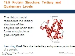
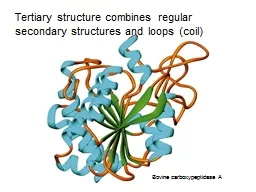
25. Complete three-dimensional shape of a given protein. Conformation.Represent the spatial relationship of the different secondary structures to one another within a polypeptide chain and how these secondary structures themselves fold into the three-dimensional form of the protein. Tertiary structure The spiral regions represent sections of the polypeptide chain that have an α-helical structure, while the broad arrows represent β-pleated sheet structures.
26. A domain is a basic structural unit within a protein molecule.Part of protein that can fold into a stable structure independently.Different domains can possess different functions.Proteins can have one to many domains depending on protein size.A polypeptide with 200 amino acids consists of two or more domains.Domains are usually connected with relatively flexible areas of protein.Protein domainPyruvate kinase (a monomeric protein): three domainsTertiary structure: Describes the relationship of different domains to one another within a protein.
27. Tertiary structure is based on various types of interactions between the side-chains of the peptide chain.
28. Stabilizing Interactions of Tertiary Structures
29. Globular ProteinsGlobular proteins fold up into compact, spherical shapes.Their functions are related to cell metabolism: biosynthesis and biodegradation, transport, catalytic function.Hydrophobic R-groups are oriented into inner part of the protein molecule, while hydrophilic R-groups are pointed towards molecule edges.Globular proteins are water soluble.
30. Example: myoglobin Globular protein that stores oxygen in musclesA single peptide chain that is mostly -helixO2 binding pocket is formed by a heme group and specific amino acid side-chains that are brought into position by the tertiary structure
31. Much or most of the polypeptide chain is parallel to a single axis Fibrous proteins are often mechanically strong and highly cross-linkedFibrous proteins are usually insoluble Usually play a structural roleFibrous proteins
32. Fibrous Proteins: KeratinsFor example, -keratins are fibrous proteins that make hair, fur, nails and skin - hair is made of twined fibrils - the -helices are held together by disulfide bonds
33. Fibrous proteins: FibroinFibroinFibroins are the silk proteins. They also form the spider websMade with a -sheet structures with Gly on one face and Ala/Ser on the otherFibroins contain repeats of [Gly-Ala-Gly-Ala-Gly-Ser-Gly-Ala-Ala-Gly-(Ser-Gly-Ala-Gly-Ala-Gly)8] The -sheet structures stack on top of each other Bulky regions with valine and tyrosine interrupt the -sheet and allow the stretchiness
34. Collagen is formed from tropocollagen subunits. The triple helix in tropocollagen is highly extended and strong. Features: Three separate polypeptide chains arranged as a left-handed helix (note that an a-helix is right-handed).3.3 residues per turn Each chain forms hydrogen bonds with the other two: STRENGTH!Nearly one residue out of three is Gly Proline content is unusually high Many modified amino acids present: 4-hydroxyproline 3-hydroxyproline 5-hydroxylysine Pro and HydroxyPro together make 30% of amino acids. Collagen amino acid composition:Fibrous proteins: Collagen is a Triple Helix
35. Covalent cross-links in collagen: alteration of mechanical properties of collagenCatalyzed by lysyl amino oxidase
36. 1. Compact protein structure Extended protein structure2. Soluble in water (or in lipid Insoluble in water (or in lipid bilayers) bilayers)3. Secondary structure is а complex Secondary structure is simplewith a mixture of a-helix, b-sheet with predominant one type onlyand loop structures 4. Quaternary structure is held Quaternary structure is usually together by noncovalent forces held together by covalent bridges5. Functions in all aspects of Structural function metabolism (enzymes, transport, (tendons, bones, muscle, immune protection, hormones, etc). ligaments, hair, skin)Globular proteins vs Fibrous proteins
37. Quaternary structure of proteinsMonomeric proteins: – built of a single polypeptide chain.Oligomeric proteins: – built of more than one polypeptide chains called subunits or monomers.
38. Quaternary structure describes the joining of two or more polypeptide subunits.The subunits each have their own tertiary structure.Bonds – non-covalent interactions.Subunits can either function independently or work co-operatively.Dissociation of a subunit results in loss of function.
39. For example: Hemoglobin A globular protein that consists of four subunits (2α and 2β, of two different types (α and β)Each subunit contains a heme group for O2 bindingBinding O2 to one heme facilitates O2 binding by other subunitsReplacement of even one amino acid in primary structure with another amino acid is critical for the function of the protein.
40. Structural organization of proteins: Summary
41.