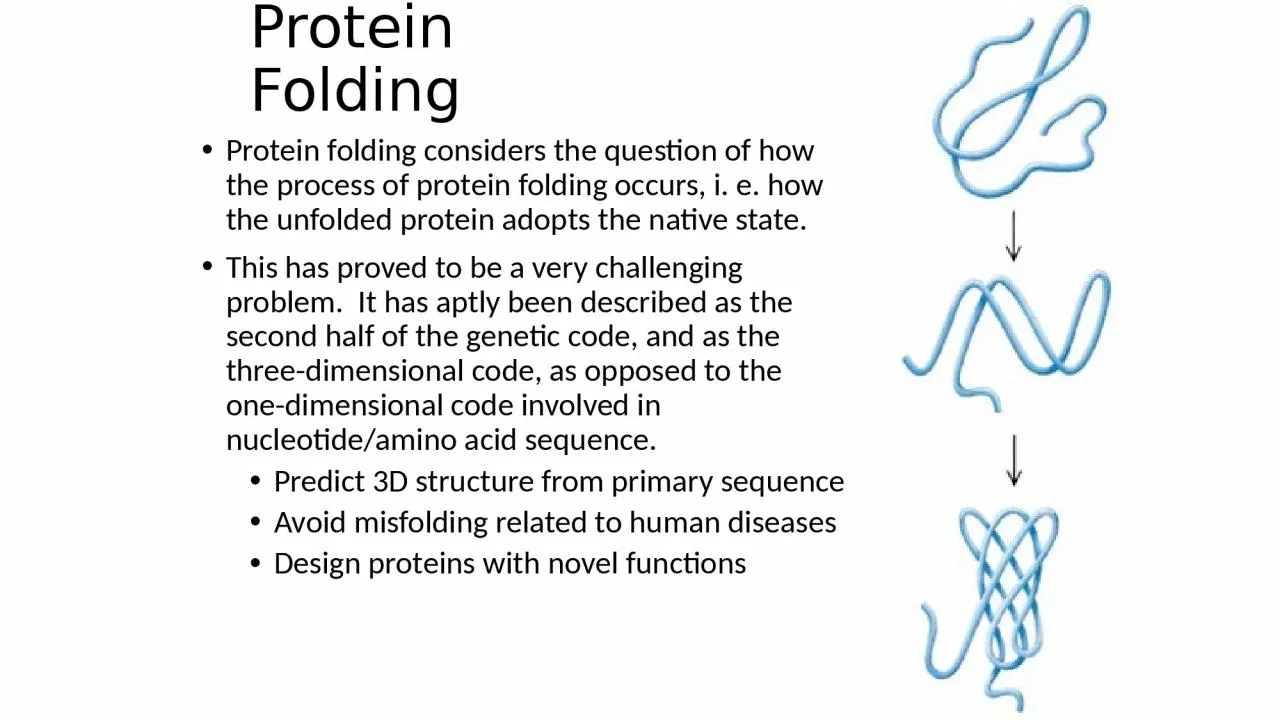
/
Protein Folding Protein folding considers the question of how the process of protein folding Protein Folding Protein folding considers the question of how the process of protein folding

Protein Folding Protein folding considers the question of how the process of protein folding - PowerPoint Presentation

white
white . @white
Follow
64 views
Uploaded On 2023-12-30

Protein Folding Protein folding considers the question of how the process of protein folding - PPT Presentation

This has proved to be a very challenging problem It has aptly been described as the second half of the genetic code and as the threedimensional code as opposed to the onedimensional code involved in nucleotideamino acid sequence ID: 1035954

protein folding native structure folding protein structure native proteins disulfide groel unfolded bonds state tertiary form groes peptide secondary

Share:

Link:

Embed:

Download Presentation from below link

Download Presentation The PPT/PDF document "Protein Folding Protein folding consider..." is the property of its rightful owner. Permission is granted to download and print the materials on this web site for personal, non-commercial use only, and to display it on your personal computer provided you do not modify the materials and that you retain all copyright notices contained in the materials. By downloading content from our website, you accept the terms of this agreement.


Presentation Transcript

1. Protein FoldingProtein folding considers the question of how the process of protein folding occurs, i. e. how the unfolded protein adopts the native state. This has proved to be a very challenging problem. It has aptly been described as the second half of the genetic code, and as the three-dimensional code, as opposed to the one-dimensional code involved in nucleotide/amino acid sequence. Predict 3D structure from primary sequenceAvoid misfolding related to human diseasesDesign proteins with novel functions

2. Anfinsen Experiment Denaturation of ribunuclease A ( 4 disulfide bonds) with 8 M Urea containing b-mercaptoethanol to random coil, no activity

3. Anfinsen ExperimentAfter renaturation, the refolded protein has native activity despite the fact that there are 105 ways to renature the protein.Conclusion: All the information necessary for folding the peptide chain into its native structure is contained in the primary amino acid sequence of the peptide.

4. Anfinsen ExperimentRemove b-mercaptoethanol only, oxidation of the sulfhydryl group, then remove urea → scrambled protein, no activityFurther addition of trace amounts of b-mercaptoethanol converts the scrambled form into native form.Conclusion: The native form of a protein has the thermodynamically most stable structure.

5. The Levinthal ParadoxThere are vastly too many different possible conformations for a protein to fold by a random search.Consider just for the peptide backbone, there are 3 conformations per amino acid in the unfolded state, For a 100 a.a. protein we have 3100 conformations.If the chain can sample 1012 conformations/sec, it takes 5 x 1035 sec (2 x 1028 year)Conclusion: Protein folding is not random, must have pathways.

6. Equilibrium Unfoldingswitch off part of the interactions in the native protein under different denaturing conditions such as chemical denaturants, low pH, high salt and high temperatureunderstand which types of native structure can be preserved by the remaining interactions

7. Equilibrium UnfoldingUsing many probes to investigate the number of transitions during unfolding and foldingFor 2-state unfolding, all probes give the same transition curves. Single domains or small proteins usually have two-state folding behavior.For 3-state unfolding, there are more than one transitions or different probes have different transition curves

8. Molten Globule State (MG)It is an intermediate of the folding transition U→MG→FIt is a compact globule, yet expanded over a native radiusNative-like secondary structure, can be measured by CD and NMR proton exchange rateIt has a slowly fluctuating tertiary structure which gives no detectable near UV CD signal and gives quenched fluorescence signal with broadened NMR chemical peaksNon-specific assembly of secondary structure and hydrophobic interactions, which allows ANS to bind and gives an enhanced ANS fluorescence MG is about a 10 % increase in size than the native state8-Anilinonaphthalene-1-sulfonic acid

9. FluorescenceA. 1 - native 3 - MG 2,4 - unfoldedB. 1 - native 3,4 - MG 2 - unfolded

10. ANS has a Strong Affinity to the Hydrophobic Surface

11. NMR of MG

12. Kinetic Folding PathwaysU→ I →II → NNot all steps have the same rate constants.Intermediates accumulate to relatively low concentrations, and always present as a mixture Identify kinetic intermediates Measuring the rate constantsFigure out the pathwaysSlow foldingFormation of disulfile bondPro isomerization

13. Unfolded StateThe unfolded state is an ensemble of a large number of molecules with different conformations.

14. MG is a Key Kinetic Intermediate

15. Three Classic Models of Protein FoldingThe Framework model proposed that local elements of native local secondary structure could form independently of tertiary structure (Kim and Baldwin). These elements would diffuse until they collided, successfully adhering and coalescing to give the tertiary structure (diffusion-collision model)(Karplus & Weaver).

16. The classic Nucleation ModelThe classic nucleation model postulated that some neighboring residues in the sequence would form native secondary structure that would act as a nucleus from which the native structure would propagate, in a stepwise manner. Thus, the tertiary structure would form as a necessary consequence of the secondary structure (Wetlaufer).

17. The hydrophobic-collapse ModelThe hydrophobic-collapse model hypothesized that a protein would collapse rapidly around its hydrophobic sidechains and then rearrange from restricted conformational space occupied by the intermediate. Here thesecondary structure would be directed by native-like tertiary structure (Ptitsyn & Kuwajima).

18. Unified Nucleation-condensation SchemeIt is unlikely that there is a single mechanism for protein folding.

19. The Folding FunnelA new view of protein folding suggested that there is no single route, but a large ensemble of structures follow a many dimensional funnel to its native structure. Progress from the top to the bottom of the funnel is accompanied by an increase in the native-like structure as folding proceeds.

20.

21. Stopped-Flow TechniqueUnfolded proteins in denaturant and buffer are placed in two syringes and mixed to allow protein folding at lower concentration of denaturants and mechanically stopped. The recording of the optical signal changes during the folding and is initiated by the macro-switch attached to the stop button.

22. Cis-trans pro

23. Folding of Cytochrome ca-helix formation is more rapid than tertiary structure rearrangements of aromatic sidechains in the folding of cytochrome c.The kinetics of these changes were determined by CD at 222 and 289 nm

24. Trapping of Disulfide-bound Intermediate The sequence of formation of disulfide bonds in proteins can be determined by trapping free cysteine residues with iodoacetate (alkylating agent). The S-carboxymethyl derivative of cysteine is stable, which be determined using chromatographic separation.

25. Structure of BPTIBovine pancreatic typsin inhibitor (BPTI) has three disulfide bonds.BPTI inhibits trypsin by inserting Lys-15 into the specificity pocket of the enzyme.

26. Folding of BPTIDisulfide bond formation was quenched at the indicated times by addition of an acid. The identities of the HPLC peaks were determined after free sulfhydryls were reacted with iodoacetate to prevent rearrangements.Only native disulfide bonds are present in the major peaks.

27. Folding of BPTIThe very fast reactions occur in milliseconds, whereas the very slow ones occur in months. The species contain 5 - 55, 14 - 38 disulfide bonds are kinetically trapped in the absence of enzymes.

28. Pulsed-labeled NMRA protein is unfolded in a D2O-denaturant solution to change amide NH groups to ND groups. Refolding is then initiated by diluting the sample in D2O to lower the concentration of denaturant. Then diluted into H2O at pH 9.0 for 10 ms and then pH 4.0. The formation of secondary and tertiary structures protects the ND group from exchange to NH. NMR is used to detect the exchanged NH groups.

29. Folding of BarnaseBarnase folds through a major pathway

30. Folding of LysozymeIn the refolding of lysozyme, the helix domain is formed before the b-sheet.Proton exchangeability was measured at different times after the initiation of folding.

31. Folding of LysozymeThe alpha helix domain is folded faster than the beta domain.

32. Parallel Pathways for the Folding of Lysozyme

33. Protein Disulfide Isomerase (PDI)The formation of correct disulfide pairings in nascent proteins is catalyzed by PDI.PDI preferentially binds with peptides that containing Cys residues. It has a broad substrate specificity for the folding of diverse disulfide-containing proteinsBy shuffling disulfide bonds, PDI enables proteins to quickly find the thermodynamically most stable pairing those that are accessible.

34. Protein Disulfide IsomerasePDI contains two Cys-Gly-His-Cys sequences. The thiols of these Cys are highly active because of their lower pKa (7.3) than most thiols in proteins (8.5), and are very active at physiological pH.PDI is especially important in accelerating disulfide inter-change in kinetically trapped folding intermediate.

35. Peptidyl Prolyl Isomerase (PPI)Peptide bonds in proteins are nearly always in the trans configuration, but X-pro peptide bonds are 6% cis.Prolyl isomerization is the rate-limiting in the folding of many proteins in vitro.PPI accelerates cis-trans isomerization more than 300 fold by twisting the peptide bond so that the C,O, and N atoms are no longer planar.

36. Peptidyl Prolyl Isomerase (PPI)

37. Molecular ChaperonesNascent polypeptides come off the ribosome and fold spontaneously, molecular chaperones are involved in their folding in vivo, and are related to heat shock proteins (hsp).The main hsp families are: "Small hsp's" - Diverse "family" 10,000 - 30,000 MW (hsp26/27 - crystallins (eye lens)) hsp40 hsp60 (e.g. GroEL in E. coli) hsp70 (DnaK in E. coli) hsp90 hsp100

38. Function of Heat Shock Proteins Minimize heat and stress damage to proteins (renaturation/degradation) Facilitate correct folding of proteins by minimizing aggregation and other misfolding Bind to nascent polypeptides to prevent premature folding Facilitate membrane translocation/import by preventing folding prior to membrane translocation Facilitate assembly/disassembly of multiprotein complexes

39. One Subunit of GroEL

40. Proteins can Fold/unfold Inside ChaperoninsA large conformational change of GroEL occurs when GroES and ATP are bound. The GroES molecule binds to one of the GroEL rings and closes off the central cavity. The GroEL ring becomes larger and the cavity inside that part of the cylinder becomes wider.

41.

42. GroES Closes Off One End of the GroEL Cylinder

43. Functional Cycle of GroEL-GroESAs shown in (a), an unfolded protein molecule (yellow) binds to one end of the GroEL-ADP complex (red) with bound GroES (green) at the other end. In (b) and (c), GroES is released from the trans-position and rebound together with ATP at the cis-position (light red) of GroEL. In (d), ATP hydrolysis occurs as the protein is folding or unfolding inside the central cavity. In (e), ATP binding and hydrolysis in the trans-position is required for release of GroES and the protein molecule. Finally, in (f), a new unfolded protein molecule can now bind to GroEL.

44.

45.